j. Soc. Cosmet. Chem., 47, 315-323 (September/October 1996) A comparison of two new in vitro phototoxicity methods to a published yeast phototoxicity method S. R. RACHUI, T. BOUFAISSAL, E. A. NEWCOMBE, and R. J. ALLEN, Stephens 6- Associates, Inc., 3310 Keller Springs Road, Suite 130, Carrollton, TX 75006 (S.R.R., T.B., E.A.N), and Shaklee Corporation, 1992 Alpine Way, Hayward, CA 94545 (R.J.A). Accepted for publication September 30, 1996. Synopsis Two new in vitro phototoxicity methods were compared to a previously published method that utilized yeast as a phototoxicity indicator. This comparison was done with eleven materials, tested in each system, to determine assay correlation. The protocols for the first two systems were similar and measured statistical differences in cell viability between irradiated and non-irradiated cell culture as a phototoxicity endpoint. The endpoint in the yeast model was a zone of inhibition generated in response to dosed phototoxic materials. The experimental sys- tems chosen for this work were (a) a monolayer of human neonatal fibroblasts and (b) the MatTek EPI-100 test system. UVA was used in these methods to elicit phototoxic responses from the test materials. It appears the MatTek EPI-100 system may be a more realistic predictor of phototoxicity as replicate standard deviations are smaller and statistically significant differences are more easily obtained. The MatTek system also has the advan- tage of being more similar to human skin than the other tested models. The monolayer system, however, may be the most sensitive of the models due to test materials directly contacting the cells and not being restricted by a stratum corneum, such as with the MatTek system. However, since statistical significance is more difficult to achieve due to greater inherent variability. The monolayer system may better be used as a screening tool. The yeast method may also be most useful when used as a rapid and inexpensive means of screening large numbers of materials prior to proceeding to more sophisticated and costly tests. INTRODUCTION Phototoxic materials are those that are not toxic under normal circumstances but that are chemically altered and become toxic when exposed to UV light. Evaluating phototoxicity, therefore, is an important addition to the toxicology profile of topically applied materials. A listing of materials that may stimulate phototoxic reactions is shown in Table I. In addi- tion, many materials that are known photoallergens may also be phototoxins. These materi- als include topical antimicrobials, fragrances, and sunscreen ingredients. Phototoxicity is normally evaluated in human subjects. This, however, can be costly and time-consuming when comparing multiple formulations. Using in vitro phototoxicity methods it is possible to evaluate materials quickly and accurately by measuring the product's effect on toxicity both before and after irradiation with UVA. UVA was se- lected as the irradiation source because most phototoxic materials respond to UVA (1). Research using a combination of UVA/UVB, such as with a solar simulator, was not done due to the strong cytotoxicity caused by UVB. This research compares two new as- says for phototoxicity evaluation to a previously published yeast method (2). 315

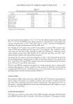
316 JOURNAL OF THE SOCIETY OF COSMETIC CHEMISTS Table I Phototoxic Drugs and Chemicals (1) Phototoxin Action Spectra Phototoxin Action Spectra Psoralens UVA Diuretics Porphyrins Visible Hydrochlorothiazide UVA Coal tar UVA Bendroflumethiazide UVA Antibiotics Furosemide Unknown Tetracyclines UVA Retinoids Fluroquinolones UVA Isotretinoin UVA/pos.UVB Nalidixic acid UVA Etretinate UVA/pos.UVB Ceftazidime Unknown Antineoplastic agents Griseofulvin UVA 5-Fluoruracil Unknown Ketoconazole Unknown Dacarbazine UVA/pos.UVB Trimethoprim Unknown Methotrexate Unknown Sulfonomides UVB Vinblastine UVB NSAIDS Dyes Arylproprionic acid derivatives Eosin Unknown Benoxaprofen UVA and UVB Fluoroscein dye Unknown Carprofen UVA Methylene blue Unknown Ibuprofen UVA Rose bengal Unknown Ketoprofen UVA Miscellaneous Nabumetone UVA Amiodarone UVA Naproxen UVA Diltiazern UVA Tiaprofenic acid UVA Fibtic acid derivatives UVB Salicylic acids Phenothiazines UVA Aspirin UVA Quinine UVA Diflunisal UVA Quinidine UVA Anthranilic acids Sulfite food derivative UVB Meclofenamic acid UVA Pyrazolidinediones Phenylbutazone UVA Oxyphenbutazone UVA MATERIALS AND METHODS MATERIALS MatTek EPI-100. The artificial tissue system selected for this study was the MatTek EPI-100. This system consists of a well-defined stratum comeurn and barrier function similar to human skin and an underlying layer of epidermal keratinocytes. Reagents used with the MatTek system were supplied by MatTek. Monolayer. A monolayer of neonatal human fibroblasts, obtained from Clonetics, was grown in a serum-free medium to approximately 80% confluence and used in the assay. Yeast. Commercial Fleischmann's yeast was selected as the indicator organism for this assay. Light source. A bank of fluorescent UVA bulbs were used for irradiation in this assay. MTT. The metabolic dye, (3-[4,5-dimethylthiazol-2-yl]-2,5-diphenyltetrazolium bro- mide) (MTT), was used as an indicator of cell viability with the MatTek EPI-100 and monolayer systems. Mitochondria within viable cells will reduce MTT from a yellow to purple color that is retained inside the cells until later extracted with isopropanol. Positive control. The positive control used in this assay was 400 I•g/ml 8-methoxypso- talin (8-MOP). This positive control was selected based on its inclusion in the pub- lished yeast method. (2-4).
Purchased for the exclusive use of nofirst nolast (unknown)
From: SCC Media Library & Resource Center (library.scconline.org)