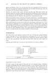
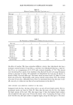
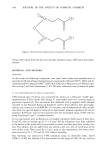
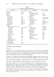
308 JOURNAL OF THE SOCIETY OF COSMETIC CHEMISTS 6e',..vA,OH O' 0 oX X OH o Figure 1. [•-Bis(carboxyethyl)germanium sesquioxide (CEG) chemical structure. thema index values from the skin spectral data obtained using a reflectance spectropho- tometer. MATERIALS AND METHODS MATERIALS In this study the following compounds were used (trade names and manufacturers in parentheses): [3-bis(carboxyethyl)germanium sesquioxide (Arlamol GEO ©, CEG) and di- methylisosorbide (Ariasolve DMI ©, DMI) (ICI Surfactants, Belgium) and [3H]water (spe- cific activity 5 mCi/ml) (Amersham, U.K.). All other substances were of analytical grade. IN VIVO PHOTOPROTECTIVE EFFECT EVALUATION UVB-induced skin erythema was monitored by means of a reflectance visible spec- trophotometer, X-Rite model 968, having 0 ø illumination and a 45 ø viewing angle, as previously reported (6). The instrument was calibrated with a supplied white standard traceable to the National Bureau of Standards' perfect white diffuser. The spectropho- tometer was connected to an IBM PS2 50 computer, which performed all color calcula- tions from the spectral data by means of the Spectrostart program supplied with the in- strument. Reflectance spectra were obtained over the wavelength range of 400-700 nm using illuminant C and 2 ø standard observer. In vivo experiments were performed on six healthy volunteers (both sexes) of skin types II and III, with an average age of 31 _ 9 years. All the volunteers were fully informed of the nature of the study and the procedures involved, and they gave their written con- sent. The subiects did not suffer from any ailments and were not on medication at the time of the study. They rested for 15 min. prior to the experiments, and room condi- tions were set at 22 4- 2oc and 40-50% relative humidity. Skin erythema was induced by UVB irradiation using an ultraviolet lamp, model UVM-57 (UVP, San Gabriel, CA), that emitted in the range of 290-320 nm, with an

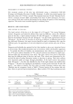
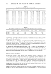
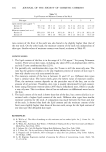
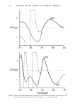
IN VIVO PHOTOPROTECTIVE EFFECT OF CEG 309 output peak at 302 nm. The flux rate measured at the skin surface was 0.80 mW/cm 2. For each subject, the minimal erythema dose (MED) was determined preliminarily, and an irradiation dose corresponding to the double of the MED was used throughout the study. For each subject, eight sites on the ventral surface of one forearm and six sites on the other were defined using a circular template (1 cm 2) and demarcated with permanent ink. Two different formulations of CEG were used: a CEG-saturated solution in dis- tilled water and a CEG-saturated solution in water:Arlasolve DMI 50:50. A solution consisting of water:Arlasolve DMI 50:50 (without CEG) was also used to assess the DMI effect on UVB-induced skin erythema. For each subject, two skin sites were left untreated but exposed to UVB radiation (control). The protocol consisted of two series of experiments (6). In the first series (pretreatment protocol), the formulations tested were applied randomly for 3 h on the skin sites of one forearm using a Hill Top chamber (Hill Top Research Inc., Cincinnati, OH) whose cot- ton pad was saturated with 150 ptl of the formulation being tested. After 3 h, the chambers were removed, the skin surfaces were gently washed with water to remove the formulation, and each pretreated site was exposed to UVB irradiation. The second series of experiments was simultaneously performed on the other forearm of the same subjects. Skin sites were exposed to UVB irradiation, and then the formula- tions tested (150 ptl) were immediately applied to the irradiated sites (using the same Hill Top chamber described above) for 6 h. After this period, the chambers and the for- mulations were removed. For both experimental protocols, UVB-induced erythema was monitored for 58 h using the reflectance spectrophotometer described above. From the skin spectral data obtained, the erythema index (E.I.) was calculated using equation 1 reported by Dawson et al. (7): 1 1 1 1 1 E.I. --- 100 [Log -- + 1.5 (Log-- + Log ) -2 (Log-- + Log )] (1) R560 R540 R580 Rs: 0 R610 where 1/R is the inverse reflectance at a specific wavelength (560, 540, 580, 510, and 610 nm). E.I. baseline values were taken at each designated site before application of the formulations tested (pretreatment protocol) or before UVB irradiation (posttreat- ment protocol), and they were subtracted from the E.I. values obtained after formula- tion application at each time point, to determine A E.I. values. For each site, the area under the response (AE.I.)-time curve (AUC) was computed using the trapezoidal rule. AUC values were inversely related to the ability of the formulations tested to inhibit UVB skin erythema. To better compare the efficacy of the different formulations tested, the percentage inhibition of UVB skin erythema (PIE) was calculated from AUC values using the following equation: AUC(c ) - AUC(T ) Inhibition % (PIE) - X 100 (2) AUC(c ) where AUC(c ) is the area under the response-time curve of sites that received no treat- ment (control), AUC(T ) is the area under the response-time curve of the sites treated with the formulations being tested. Statistical analysis of the results were performed us- ing Student's t-test.
Purchased for the exclusive use of nofirst nolast (unknown)
From: SCC Media Library & Resource Center (library.scconline.org)