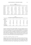
NEW PHOTOTOXICITY METHODS 317 METHODS &ITT com12•tiai/ity. Cell viability, the endpoint for phototoxicity with the MatTek EPI- 100 and monolayer systems, was measured using MTT. In some cases, test materials are capable of reducing MTT and causing its color change from yellow to purple, which would normally be indicative of cellular metabolic activity. This is controlled by com- bining equal volumes of test materials and MTT and incubating them in the dark for a time equal to that used for MTT exposure in the assay. At the end of the incubation period, materials that have not caused a color change to purple are considered MTT- compatible, while materials that have caused a color change to purple are incompatible. Further steps are taken for MTT-incompatible materials to ensure these materials do not cause false readings in the assay. None of the test materials used in this study were MTT-incompatible. In the monolayer and MatTek EPI-100 systems, materials that show a significant (p • 0.05) increase in toxicity after UVA irradiation, when compared to the toxicity of the same materials without irradiation, are considered phototoxic. Statistical signifi- cance is important in the interpretation of results, as statistical measurements effectively account for biologic variability. Results that directionally appear to indicate phototoxic- ity may be attributable to biologic variability unless the difference between treatment and no treatment is large enough to preclude the effects of biologic variability. Al- though statistical significance is a more objective endpoint for measuring phototoxicity, Duffy et•/. have established a protocol in which the cutoff for phototoxicity is based on the magnitude of difference in viability between non-irradiated and irradiated cell cul- ture (5). &Io,o/ayer. Since test materials cannot easily be assayed neat in monolayer systems, test materials were diluted to 1% of their stock concentration. Dilution of test materials also served to avoid cytotoxicity, which was noted with several of the materials at higher concentrations in previous assays with monolayer culture. Fibroblasts were suspended in Dulbecco's Modified Eagles medium with 10% fetal bovine serum (DMEM). One (1) ml aliquots of this mixture were then seeded into wells of 24-well plates at a titer of 2 X 10 4 cells/well. The cells were allowed to fix to the plate for six hours, and then fresh DMEM containing test material, was added in tripli- cate using duplicate plate sets, one set for UVA exposure and one set that was not ex- posed to UVA. The final volume of each well was 1.5 ml, with the final concentration of test material being 1% of the neat concentration. Once dosed, both plate sets were in- cubated for 24 hours at 37øC and 5% CO 2 to allow for material metabolism. After 24 hours' incubation, the set for UVA irradiation was removed from incubation and placed under a bank of UVA bulbs at approximately 1.5 mw/cm 2 for 30 minutes, resulting in a UVA dose of about 3 joules. After irradiation, the plates were placed back into the incu- bator with the non-irradiated plates for another 24-hour period. After this second 24- hour incubation, both plate sets were removed from the incubator and the DMEM was aspirated and replaced with 1 mg/ml MTT prepared in DMEM. The plates were incu- bated at 37øC and 5% CO 2 in the presence of MTT for two hours. At the end of this period, the MTT solution was aspirated, the cells were rinsed with PBS, and 1 ml of isopropanol was added to each well. The plates were incubated at room temperature for one hour to extract the converted MTT from the cells. At the end of incubation, the ab- sorbance of each well was measured at 570 nm. The viability of the culture was deter- mined using the following formula:

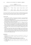
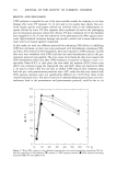
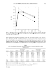
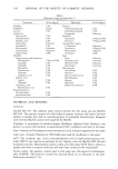
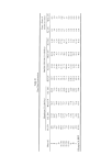
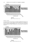
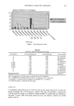
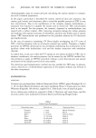
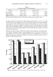
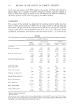
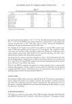
318 JOURNAL OF THE SOCIETY OF COSMETIC CHEMISTS Mean absorbance density from cultures treated with test material X 100 = %viability Mean absorbance from untreated control cultures MatTek EPI-100. The MatTek tissue was transferred from its shipping tray to six-well plates containing 0.9 ml culture media. Tissue was dosed with 100 ptl of test material in triplicate using duplicate plate sets, one set for UVA exposure and one set that was not exposed to UVA. Test materials were dosed neat in this assay by applying neat ma- terials to the stratum corneum of the tissue construct. Once dosed, both plate sets were incubated at 37øC and 5% CO 2 for 24 hours to allow for material penetration and me- tabolism. After 24 hours' incubation, the set for UVA irradiation was removed from in- cubation and placed under a bank of UVA bulbs at approximately 1.6 mw/cm 2 for 30 minutes, resulting in a UVA dose of about 3 joules. After irradiation, the plates were placed back into the incubator with the non-irradiated plates for another 24-hour pe- riod. After this second incubation, both sets of plates were removed from the incubator, and the tissue was rinsed free of test material with PBS and transferred to a 24-well plate containing 300 ptl MTT. At the end of three hours, the MTT solution was aspi- rated, the cells were rinsed with PBS, and 1 ml of isopropanol was added to each well. The plates were incubated at room temperature for two hours to extract the converted MTT from the cells. At the end of incubation, the absorbance of each well was mea- sured at 570 nm. The viability of the culture was determined using the following for- mula: Mean absorbance from cultures treated with test material X 100 = % viability Mean absorbance from untreated control cultures Yeast. A culture of commercial Fleischmann's yeast was prepared by adding 200 mg of yeast to 100 ml of deionized water. This suspension was then diluted by adding 7.4 ml of the suspension to 92.6 ml of deionized water. Cultures were made by generously streaking this prepared culture onto Sabouraud Dextrose agar in six-well plates. Test material was exposed in triplicate using duplicate sets, one set for UVA exposure and one set that was not exposed to UVA. Test materials were dosed neat in this assay by applying them to circular filter pads and allowing them to dry for 15 minutes. Once dry, the filter pad was placed onto the center of the yeast culture. The plate set for UVA exposure was then placed under a bank of UVA bulbs and irradiated overnight at 0.8 mw/cm 2, while the non-irradiated plate set was placed in the dark at room temper- ature and incubated overnight. The following day, the irradiated plates were removed from UVA exposure and placed in the dark with the non-irradiated set of plates for an- other overnight period. On the third day, plates were examined for a zone of inhibition surrounding the filter disk. A material that causes a zone of inhibition of greater than 2 mm in diameter around the filter disk, when compared to the non-irradiated con- trols, is considered phototoxic. The zones of inhibition were measured with calipers (2). RESULTS AND CONCLUSIONS Phototoxicity in the monolayer and MatTek EPI-100 assays is noted when a significant (p -- 0.05) difference is seen between irradiated and non-irradiated cultures. The re- suits for all three test systems are listed in Table II and presented graphically in Fig- ures 1-3.
Purchased for the exclusive use of nofirst nolast (unknown)
From: SCC Media Library & Resource Center (library.scconline.org)