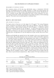
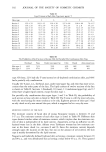
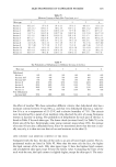
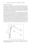
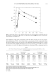
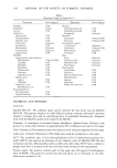
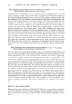
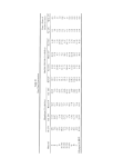
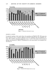
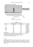
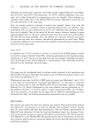
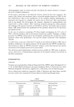
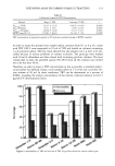
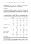
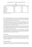
NEW PHOTOTOXICITY METHODS 32 ! Zone of inhibition in millimeters 10 8 6 4 2 O •n0:uv treatment r-•with UV treatment 48 hours Fiõur½ 3. Yeast phototoxicity assay. Table III Statistical Analysis of Phototoxicity Results Test material Monolayer fibroblasts MatTek EPI-100 Yeast method • A NST NST Not phototoxic B NST NST Not phototoxic C NST NST Phototoxic D NST NST Not phototoxic E (1%) NST NST Not phototoxic E (3%) NST NST Phototoxic E (5%) NST NST Not phototoxic F (1%) NST NST Not phototoxic G NST a NST Not phototoxic H NST a NST Not phototoxic I NST a NST Not phototoxic 400 •g/ml 8-MOP NST STN Phototoxic NST: No significant (p • 0.05) increase in toxicity due to UVA irradiation. STN: A significant (p • 0.05) increase in toxicity was noted due to UVA irradation. •A significant (p • 0.05) increase in viability was noted after UVA irradiation. b No statistical analysis was done for the yeast assay. MONOLAYER No significant phototoxicity (p • 0.05) was seen in this assay with any of the test ma- terials, including the 8-MOP positive control. The lack of significant phototoxicity with the positive control is attributed to high standard deviations that are inherent in monolayer systems. HTe directional results, however, do indicate phototoxicity of the positive control.

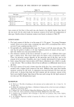
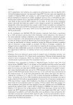
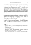
322 JOURNAL OF THE SOCIETY OF COSMETIC CHEMISTS Although not statistically significant, directional trends suggest Material C and Mater- ial E at 0.01% and 0.03% were phototoxic. At 0.05%, however, Material E was cyto- toxic, and reliable phototoxicity measurements were not possible. These findings are consistent with trends seen in the MatTek EPI-100 system. Materials C and E (at 3%) were also phototoxic in the yeast assay. Since no stratum corneum is present to restrict test material contact with cells, the monolayer method is a sensitive means of assessing phototoxicity. In addition, since mammalian cells are used, phototoxicity of the test compound and potential metabo- lites can be assessed. Due to the nature of the test system, however, materials cannot easily be assayed neat. In this test, cytotoxicity was more of an issue than with the Mat- Tek EPI-100 and yeast methods. Also, the absence of a stratum corneum and barrier function does not allow for a realistic evaluation of phototoxicity on intact skin. While no statistical differences were noted with the materials, directional data correlated with the results in the yeast assay. MatTek EPI- 100 A significant (p • 0.05) increase in toxicity as a result of the 8-MOP positive control was noted in tissue irradiated with UVA, indicating phototoxicity. No significant pho- totoxicity (p • 0.05) was seen in this assay with any test material. Although not signif- icant, directional trends indicate Material C was phototoxic. This directional trend was also noted in the monolayer and yeast assays. YEAST This assay was the benchmark used to evaluate results obtained in the monolayer and MatTek EPI-100 assays. Materials that caused a zone of inhibition of greater than 2 mm were defined as phototoxic (2-4). Phototoxicity was noted with the 8-MOP positive control and Materials C and E (3%) in this assay. The phototoxicity associated with Material C in this assay is consistent with the trends noted in both the MatTek EPI-100 and monolayer systems. Although Material E at 3% showed phototoxicity, the same material was not phototoxic at 1%. The difference between 1% and 3% may be a simple concentration effect. At 5 %, Mate- rial E was cytotoxic, as indicated by the large zone of inhibition surrounding the non- irradiated filter disk, and reliable phototoxicity assessments were not possible. DISCUSSION The results seen across the three test systems were similar. Directional trends indicate that Material C was phototoxic in both the MatTek and monolayer systems. Results with the yeast model confirmed this phototoxicity. The two lowest concentrations of Material E were phototoxic in both the yeast and monolayer assays, but, because of cyto- toxicity, phototoxicity was not noted at the highest concentration. The lack of response with Material E in the MatTek EPI-100 system may be due to the presence of the stra- tum corneum, which serves as a protective barrier and limits material penetration to the underlying epidermis.
Purchased for the exclusive use of nofirst nolast (unknown)
From: SCC Media Library & Resource Center (library.scconline.org)