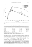
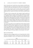
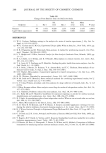
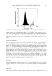
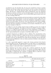
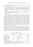
304 JOURNAL OF THE SOCIETY OF COSMETIC CHEMISTS Table I The Results of the Ratio of Wrinkle Calculated by Image Analysis Treatment Ratio of wrinkle (image analysis %) + S.D. Original a Artificial b Control 1.74 + 0.80',-• 0.56 + 0.43*,%:• UV-B only 21.68 +_ 2.06*,%•c 2.12 + 1.32*,%•c Temporary groove only • 3.81 + 1.89',? 4.32 + 1.30' Temporary groove after UV-B d 14.05 + 5.35:1: 5.68 + 3.70* UV-B after temporary groove e 16.91 + 4.58•c 13.74 + 4.07',•c * Significantly different from the group characterized by production of temporary groove after UV-B irradiation (p 0.01). ? Significantly different from the group characterized by UV-B irradiation after production of temporary wrinkle (p 0.01-0.05). • Significantly different from the group characterized only by production of temporary wrinkle (p 0.01-0.05). a Original wrinkle at right angles with the midline, which was observed in usual state. b Artificial wrinkle parallel to the midline, which was unusual direction of wrinkle formation. c Group characterized by production of temporary wrinkle without UV-B irradiation. d Group characterized by production of temporary wrinkle after UV-B irradiation. e Group characterized by UV-B irradiation after production of temporary wrinkle. before--production of the temporary groove are necessary for wrinkle formation in this mouse model, and we suggest that the skin morphology at the time of UV-B irradiation is important for wrinkle formation. Recently, wrinkle development in these mice in response to irradiation has been taken to be an indicator of chronic UV light exposure, or photoaging (4-7). Bissett et aL (8) reported slight wrinkle formation after five weeks of UV-B irradiation and permanent wrinkles after about 15 weeks. Kiss et aL (11) investigated the effects of high- and low-dose UV-B exposure on the degree of hairless mouse skin wrinkling and observed that wrinkle formation does not parallel the UV-B irradiation dose. Moloney et aL (12) reported that visible signs of wrinkling were present after approximately six weeks of UV-B irradiation and were very apparent after ten weeks of irradiation. The direction of wrinkle formation observed in our study differed from that in the above studies. How- ever, the timing of the initiation of wrinkles seemed to be similar. This observation may suggest that the total cumulative dose is important for the timing of the initiation of wrinkles. Previously, we considered two possible mechanisms for wrinkle formation after tempo- rary fixation. Tissue destruction and subsequent tissue reconstruction occur in the areas exposed to UV-B but not in other areas, and this may cause distortion in the tissue, resulting in wrinkle fixation. Alternatively, when reconstruction after destruction is continuously accelerated in the entire tissue exposed to UV-B, tissue reconstruction may proceed along the temporary groove, causing fixation of the wrinkle. In our experimental system, the temporary groove was confirmed to be restored to the previous state after one to two hours. In this study, no artificial wrinkling was induced by production of an artificial temporary groove after UV-B irradiation, and these findings suggest that the latter hypothesis is unlikely. Therefore, the primary cause of wrinkle formation after fixation followed by UV-B irradiation may be differences in local changes due to uneven

SKIN MORPHOLOGY AND WRINKLE FORMATION 305 UV-B irradiation. On the other hand, our results show a slight delay in the formation of longitudinal wrinkles after temporary fixation, compared with the formation of original transverse wrinkles. Thus, there remains a slight possibility that the latter hypothesis described above may explain wrinkle formation. The connective tissue elements collagen and elastin are known to be degraded by collagenase and elastase produced by inflammatory cells (14,15) and fibroblasts (16,17). Collagenase mRNA levels in the dermis (18) and cultured fibroblasts (19) were reported to increase after UV-A irradiation. It is well known that exposure of keratinocytes in culture (20) or of the skin in vivo (21,22) to UV irradiation induces marked elevation of release of several cytokines, including interleukin-1 (IL-1) and tumor necrosis factor (TNF). UV-inducible IL-1 has also been shown to stimulate the synthesis of elastase (23) and to increase collagenase activity (24) in normal human fibroblasts. On the other hand, recent studies have shown up-regulation of the elastin mRNA level by IL-i[• (25) and its down-regulation by TNF (26). Thus, it is conceivable that the synthesis and degra- dation of connective tissue elements are regulated by cytokines. In addition, recent studies have shown the expression of collagenase by normal human fibroblasts in re- sponse to singlet oxygen radicals generated by UV-A irradiation (27,28). Active oxygen species induce cross-linkage of collagen in vitro (29) and in vivo (30). Oxidative autoac- tivation of latent collagenase by human neutrophils has also been reported (32). Appli- cation of a radical scavenger has been reported to inhibit wrinkle formation in animals (31). Considering the expression of proteins such as collagenase, there may be a time lag between UV-B exposure and expression of enzyme activity, supporting the uneven irradiation theory. However, cross-linking due to active oxygen species and activation of latent-type collagenase (32) suggests that the skin morphology at the time of UV-B irradiation is important for wrinkle formation. Although it is unclear which mechanism is involved, wrinkles can be formed in any direction by temporary fixation followed by UV-B irradiation. Further studies are needed to determine whether this phenomenon occurs in other animal species and whether uneven irradiation, such as that by slit rays, is also the same effect. REFERENCES (1) A.M. Kligman, P. Zheng, and R.M. Lavker, The anatomy and pathogenesis of wrinkles, Br. J. Dermato/., 113, 37•42 (1985). (2) T. Tuji, T. Yorifuji, Y. Hayashi, and T. Hamada, Light and scanning electron microscopic studies on wrinkles in aged persons' skin, Br. J. Dermato/., 114, 329-335 (1986). (3) G. E. Pierad and C. M. Lapiere, The microanatomical basis of facial frown lines, Arch. Dermato/., 125, 1090-1092 (1989). (4) H. M. Daniell, A study in the epidemiology of "crow's feet," Ann. Intern. Med., 75,873-880 (1971). (5) G.L. Grove, M.J. Grove, and J. J. Leyden, Optical profilometry: An objective method for quantifi- cation of facial wrinkles, J. Am. Acad. Dermato/., 21, 631-637 (1989). (6) C. E. Griffiths, T. S. Wang, T. A. Hamilton, and J. J. Voorhees, A photonumeric scale for the assess- ment of cutaneous photodamage, Arch. Dermato/., 128, 347-351 (1992). (7) P. Corcuff, J. Rigal, andJ. L. Leveque, Skin relief and aging,J. Soc Cosmet. Chem., 34, 177-190 (1983). (8) D.L. Bissett, D.P. Hannon, and T.W. Orr, An animal model of solar-aged skin: Histological, physical, and visible changes in UV-irradiated hairless mouse skin, Photochem. Photobio/., 46, 367-378 (1987). (9) D. L. Bissett, D. P. Hannon, and T. W. Orr, Wavelength dependence of histological, physical, and visible changes in chronically UV-irradiated hairless mouse skin, Photochem. Photobio/., 50, 763-769 (1989).
Purchased for the exclusive use of nofirst nolast (unknown)
From: SCC Media Library & Resource Center (library.scconline.org)