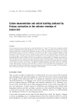
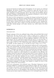
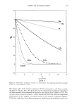
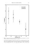
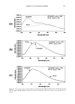
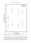
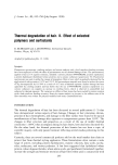
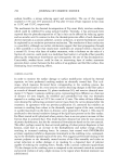
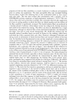
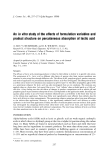
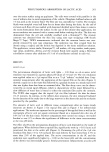
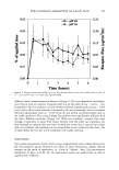
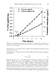
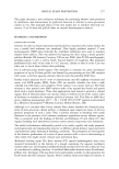
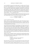
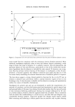
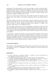
266 JOURNAL OF COSMETIC SCIENCE • 2 T 2 gl finite dose 75 •tl infinite dose 0 I I I 0 1 2 3 4 5 6 Time (hours) Figure 5. Time dependency of transdermal penetration of lactic acid delivered from a 2-pl finite-dose o/w emulsion film (n = 6) and a 75-pl o/w infinite dose (n = 7) at pH = 3.8 as assessed by the cumulative absorption in the receptor phase. Error bars omitted for clarity. These penetration results suggest that the penetration pathways for lactic acid in the corneum for the topical-film and infinite-dose applications might be different. In the finite-dose situation, the SC, except for a short (ca 20 minutes) period following appli- cation, was not hydrated. Thus the active has to penetrate the corneum through a hydrophobic (possibly the lipid) pathway. At pH 3.8, lactic acid is 50% ionized (pK a = 3.8), whereas at pH 7.0, it is 99.9% ionized. It is known that charged species penetrate poorly through the hydrophobic lipid bilayers. These results are in agreement with in vitro measurements by Michaels et al. (20), which suggest that the permeabilities of the ionized forms were -1/20 of those for their un-ionized forms. Assuming that stratum corneum is the principal barrier to lactic acid penetration, the permeability coefficient, P, can be calculated from the receptor flux, J, from the fol- lowing equations (22):

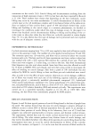
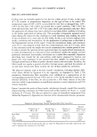
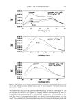
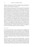
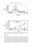
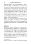
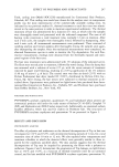
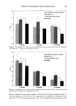
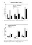
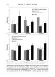
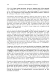
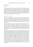
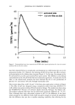
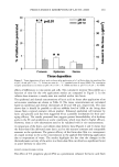
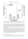
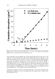
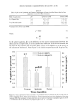
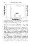
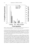
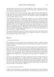
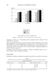
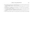
PERCUTANEOUS ABSORPTION OF LACTIC ACID 267 Table II Effect of pH on the Epidermal and Dermal Concentration of Lactic Acid Six Hours After In Vitro Application of an Oil-inWater Emulsion Mode of application pH Epidermis (mM) Dermis (mM) 2plTopicalfilm 3.8 24.9ñ3.5* 1.6ñ0.3 7.0 4.6ñ1.1 1.1ñ0.1 75plOccludedpatch 3.8 23.1ñ5.6 14.1ñ4.5 7.0 20.4ñ4.5 5.4ñ2.2 * Standard error of the mean (SEM). j=PAC (1) where KD p _ (2) L In the above equations, AC is the difference in the active concentrations between the donor and the receptor sides, K is the distribution coefficient of the active between the top layer of the corneum and the donor phase, and D is the diffusivity of the active in the corneum of thickness L. From Figure 4, the initial receptor flux was 0.16 pg/cm2/hr, 0.9 0.8 0.7 0.6 0.5 0.4 0.3 0.2 0.1 • 75 gl o/w with PG •e l* {--I 75 gl o/w without PG pO.01 T 8O 0.0 0 Corneum Epidermis Dermis Tissue deposition Figure 6. Tissue deposition of lactic acid from a 75-1•1 infinite dose o/w emulsion at pH 3.8 with (5%) or without propylene glycol. Tissue concentrations (n = 6) were measured six hours after application. Error bars represent SEM. The cumulative receptor penetration at six hours in the presence of propylene glycol is 0.2% ñ 0.0 SEM and that in the abscence of propylene glycol is 0.1% ñ 0.0 SEM. 70 6O • 5O m 4O • 30 20 lO '
Purchased for the exclusive use of nofirst nolast (unknown)
From: SCC Media Library & Resource Center (library.scconline.org)