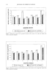
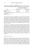
342 JOURNAL OF COSMETIC SCIENCE O II I H2CO - P--O I / o- I OH Figure 1. Schematic representation of a cyclobutane pyrimidine dimer. UV-induced melanogenesis or tanning is a natural protective response of the skin: an increase in intracellular melanin reduces ultraviolet transmission (5). In addition, enzy- matic repair systems are available to remove cellular damage inflicted by UV, whereas the skin's immune system destroys possible precancerous cells. Tanning is generally considered a natural sunscreen, but its underlying mechanisms have not yet been fully elucidated. Melanin is biochemically synthesized from tyrosine. Epi- cutaneous application of a mixture of tyrosine/adenosinephosphates/riboflavin before UV exposure was shown to increase and accelerate the tanning response (6). Interestingly, UV-induced DNA damage and/or its repair have been linked to melanogenesis (7-9). In the present study we have investigated the effect of the epicutaneous application of tanning accelerators (Unipertan © VEG-2002) on the level of DNA damage in epidermal cells at various time points after exposure to UV. MATERIAL AND METHODS TANNING ACCELERATION Unipertan © VEG-2002 (international patents by Induchem, Switzerland) was used as a tanning accelerator, containing tyrosine (8%), adenosine phosphates (1%), riboflavin (0.3%), vegetable protein hydrolysate, and butylene glycol (20%). This complex of ingredients has been shown to be effective in tanning acceleration (6). Two l•l/cm 2 of a 5% solution of Unipertan © VEG-2002 in water was applied to human skin, in line with recommendations by the FDA and COLIPA for the testing of sunscreens. IN VITRO STUDY Human abdominal skin was obtained directly after surgery from three female donors at

DNA DAMAGE AND REPAIR 343 the academic Hospital of Utrecht (The Netherlands). Skin discs (8-mm diameter) were prepared and cultured on a microporous membrane as described by Van de Sandt and Rutten (10). A one-microliter mixture of tanning enhancers (5% Unipertan © VEG-2002 in sterile water) was brought onto the skin discs using a positive displacement pipette. The substance was evenly spread over the skin area with a disposable spatula (Poly- Pipets, Inc., Englewood Cliffs, NJ). Incubation with the substance was performed in a humidified incubator (37øC, 5% CO2) the skin discs were exposed to UVB (0, 3000 J/m 2) from Westinghouse FS20 sunlamps (2 x 20 W). The UV-dose rate was 0.37 mW/cm 2 as determined with a UVX dosimeter (UV Products, San Gabriel, CA) equipped with a UVX-31 sensor for the measurement of UVB. At various time points after exposure to UV radiation (0, 6, 24 h) the skin was fixed with phosphate-buffered 4% formaldehyde and paraffin sections were prepared. CLINICAL TRIAL Besides the studies with human skin organ cultures, a clinical trial was performed in which three volunteers participated, two males and one female, with written informed consent. At first, each of the volunteer's personal minimal erythema dose (MED) was assessed using Westinghouse FS20T12 sunlamps (4x). The doses that resulted in a just perceptible redness were 1100 J/m 2 (volunteer 1), 1400 J/m 2 (volunteer 2), and 800 J/m 2 (volunteer 3). Two 121/cm 2 of a 5% solution of Unipertan © VEG-2002 was topically applied to one of the arms of each volunteer. After 1 h incubation the arm was irradiated with 3 MED, and punch biopsies were taken immediately after and at 24 h after irradiation an additional unexposed skin biopsy was obtained from the same treated arm. The other arm was treated with UV only, and two skin biopsies (t = 0, 24 h) were taken. The skin samples were immediately transferred to a fixative comprised of phos- phate-buffered 4% formaldehyde, and paraffin sections were prepared. IMMUNOSTAINING OF UV-DNA DAMAGE Paraffin sections were deparaffinized by subsequent 2-min incubations in xylene (2 x), ethanol 100% (2x), ethanol 96%, ethanol 70%, and PBS. The slides were then boiled for 10 min in 10 mM citrate buffer (pH 6.0), rinsed with PBS (2x), and used for immunostaining. The skin sections were stained with monoclonal antibodies against cyclobutane pyrimidine dimers [hybridoma clone H3 (11)] and with goat-anti-mouse- IgG fluorescein-labeled secondary antibodies. The nuclei of the skin cells were coun- terstained with propidiumiodide. Nuclear green fluorescence in the epidermal cells proportional to the level of pyrimidine dimers was assessed with a scanning laser mi- croscope (Zeiss LSM-41, Oberkochen, Germany) using image processing and image analysis. The procedure for the immunostaining and measurement of the fluorescence has been described (11,12). RESULTS IN VITRO STUDY Skin organ cultures from three donors were used. Irradiation of skin discs with 3000
Purchased for the exclusive use of nofirst nolast (unknown)
From: SCC Media Library & Resource Center (library.scconline.org)