j. Cosmet. Sci., 50, 363-385 (November/December 1999) Structural characteristics and permeability properties of the human nail: A review GOURI V. GUPCHUP and JOEL L. ZATZ, College of Pharmacy, Rutgers University, 160 Frelinghuysen Road, Piscataway, NJ 08854. Accepted for publication October 15, 1999. Synopsis The human nail forms a resistant barrier to the topical penetration of actives. Thus, treatment of nail disorders, such as fungal infections, remains a challenge because of the difficulty encountered in achieving therapeutic concentrations of drugs at the site of infection. The nail is primarily composed of a highly cross-linked keratin network that contains several disulfide linkages. This unique structure results in a highly effective permeability barrier. Nail penetration has been reported to be affected by molecular size and hydrophilicity smaller, water-soluble molecules are found to preferentially permeate the nail. Permeation of undissociated drugs is favored in certain instances. Also, some studies indicate that the nature of the vehicle can influence drug penetration. Recent research has focused on improvement of penetration of topically applied actives into and through the nail. Studies have shown that compounds containing sulf- hydryl groups in conjunction with keratolytic agents can significantly enhance drug penetration, relative to a control formulation (without enhancer). Such sulfhydryl compounds are thought to reduce the disulfide linkages in the nail keratin matrix. Thus, although some success has been achieved in enhancing penetration of drugs through the nail, further research is required to achieve successful topical products for treatment of nail infections. INTRODUCTION The human nail acts as a protective covering to the delicate terminal phalanges of the fingers and toes and helps in grasping small objects. Changes in the appearance of the nail result from a variety of conditions such as fungal, bacterial, and viral infections or dermatological disorders (1-3). Various cosmetic procedures such as application of ar- tificial acrylic nails, use of nail hardeners, and manicures can also result in nail disorders (4). Fungal infections of the nail, called onychomycosis (OM), are some of the most com- monly encountered dermatological disorders, typically manifested as localized infections of the nail or nail bed. OM has widespread incidence and is thought to account for 40% of all nail disorders. It is estimated that 4.9-12.3 million people are affected with OM in the United States. The incidence of OM increases with age, and toenails are infected about seven times more frequently than fingernails. Mycotic nail infections are caused by dermatophytes, yeasts, and nondermatophyte molds, although dermatophytes are be- lieved to be the principal causative organisms in OM (3,5). Symptoms of OM include 363

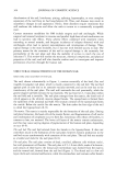
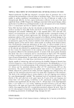
364 JOURNAL OF COSMETIC SCIENCE discoloration of the nail, brittleness, pitting, splitting, hypertrophy, or even complete separation of the nail from its bed (onycholysis) (6). Thus, nail diseases may result in unaesthetic changes in nail appearance. Hence, these disorders require treatments that will eradicate the infection and allow the nails to return to a cosmetically acceptable state. Current treatment modalities for OM include surgery and oral antifungals. While surgical nail removal (avulsion) is invasive and painful, high doses of oral medication can lead to systemic side effects. These adverse effects combined with treatment times extending to several months, and frequent incidences of relapses observed with oral antifungals, often lead to patient noncompliance and interruption of therapy. Thus, topical therapy is the most desirable, but it has met with limited success to date. The primary reason for the resistance of the nail to topical therapy is the extremely low permeability of the nail plate and thus the inability of actives to reach the site of infection (7). This review will outline the structure, chemical composition, and physical properties of the nail, and will also describe studies used to investigate and improve permeation of actives through the human nail. STRUCTURAL CHARACTERISTICS OF THE HUMAN NAIL STRUCTURE AND ANATOMY OF THE NAIL The nail, shown schematically in Figure 1, consists essentially of the hard, fiat, and roughly rectangular nail plate, which is closely connected to the nail bed. The nail bed appears pink in color due to its extensive vascular network, and can be seen due to the translucency of the nail plate. The nail wall surrounds the nail proximally, while the groove-shaped nail fold encloses the nail laterally. The nail root lies 3-5 mm deep within the nail fold and is invisible. The nail plate emerges from the matrix, the distal end of which appears as the whitish crescent-shaped lunula. The eponychium is formed from the epidermis of the proximal nail wall. The stratum corneum of the eponychium forms the cuticle. Below the cuticle lies the matrix. The skin under the free edge of the nail is called the hyponychium (8-9). The matrix. The matrix is mainly responsible for the formation of the nail plate. During nail plate formation, the basal cells of the matrix fiatten, and fragmentation of cell nuclei and condensation of cytoplasm occur to form fiat, keratinous cells whose cell borders, in contrast to hair, are retained. The lower cell layers of the matrix contain melanocytes, and this may cause varying degrees of pigmentation of the human nail plate, depending on race (8-9). The nail bed. The nail bed extends from the lunula to the hyponychium. It does not contribute much to the formation of the nail plate however, keratin production in the nail bed occurs synchronously with extension of the nail plate. The nail bed acts mainly as a holder and slide for the nail plate (8-9). The nail plate. The nail plate consists of dead, cornified, adherent cells without nuclei, but with prominent cell borders. The nail plate is 0.5-1.0 mm thick, made of o•-keratin, and consists of three layers: the dorsal and intermediate nail, formed from the matrix and the ventral nail, formed from the nail bed (Figure 1). The dorsal nail is a few cell layers thick and contains hard keratin, while the intermediate nail contains softer keratin
Purchased for the exclusive use of nofirst nolast (unknown)
From: SCC Media Library & Resource Center (library.scconline.org)