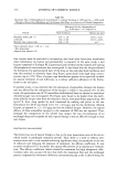
STRUCTURE AND PERMEABILITY OF HUMAN NAIL 367 Table I Amino Acid Composition (as residues per 100 residues) of Human Nail, Human Hair, and Human Stratum Corneum, Compared With That of Sheep Wool, Horn, and Hoof Human Sheep Stratum Amino acid Nail Hair corneum Wool Horn Hoof Lysine 3.1 2.5 4.2 2.7 3.8 4.0 Histidine 1.0 0.9 1.5 0.8 1.3 0.9 Arginine 6.4 6.5 3.8 6.2 6.7 7.2 Asparric acid 7.0 5.4 7.9 5.9 7.8 8.4 Threonine 6.1 7.6 3.0 6.5 4.8 5.0 Serine 11.3 12.2 13.6 10.8 9.6 9.5 Glutamic acid 13.6 12.2 12.6 11.1 12.9 13.7 Proline 5.9 8.4 3.0 6.6 3.8 4.0 Glycine 7.9 5.8 24.5 8.6 11.1 9.1 Alanine 5.5 4.3 4.4 5.2 5.9 6.4 Valine 4.2 5.5 3.0 5.7 5.2 5.7 Methionine 0.7 0.5 1.1 0.5 0.8 0.8 Isoleucine 2.7 2.3 2.7 3.0 3.3 3.6 Leucine 8.3 6.1 6.9 7.2 9.1 9.5 Tyrosine 3.2 2.2 3.4 3.8 5.0 4.0 Phenylalanine 2.5 1.7 3.2 2.5 2.7 2.7 Hal f-cys tine 10.6 15.9 1.2 13.1 6.2 5.7 Sulfur (%) 3.2 4.5 1.4 3.8 2.1 2.2 Adapted from references 13, 14. position of human nails differs considerably from that of horn or hoof derived from sheep. In general, significant amounts of glutamic acid, half-cystine, arginine, aspartic acid, serine, and leucine are present in human nails (9,13-15). The nail cellular envelope contains large amounts of proline, unlike the cell matrix, which contains numerous cysteinyl residues (16). Carbohydrates are also found to be present in the cell membrane complexes of human nails. Allen eta/. (17) demonstrated by formic acid extraction and lectin-binding studies that sugar characteristics of membrane glycoproteins (i.e., man- nose, galactose, N-acetylglucosamine, and N-acetylgalactosamine) were present in the cell membrane complexes of nails. The lipid content of nails is less than 5% (8-9), while the sulfur content is high, as in hair (Table I) (13-4). Nitrogen is another major component, resulting from the presence of high levels of keratin. The nail also contains low levels of Ca, Mg, Na, K, Fe, Cu, and Zn (18). The calcium content was earlier thought to be responsible for the hardness of the dorsal nail (19). However, it is now believed that the presence of disulfide linkages, rather than the calcium content, is responsible for nail hardness (8-9). Trace amounts of Cr, Se, Au, Hg, Ag, and Co have also been reported in human thumbnails (20). STRUCTURE OF KERATIN The main form of keratin present in all mammalian hairs, wool, horns, claws, nails, and quills is tx-keratin. The presence of covalent disulfide cross-links, which occur due to the high proportions of cystine, confers an exceptional degree of physical and chemical stability on the keratins. In addition to the intermittent disulfide cross-links, there are

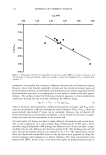
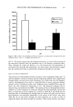
368 JOURNAL OF COSMETIC SCIENCE other secondary bonds also present in the keratin structure. These include Van der Waals interactions, hydrogen bonds, and coulombic interactions. The coulombic interactions arise because of the presence of negatively charged carboxylic acid groups (COO-) and positively charged amino groups (NH3 +) formed on the side chains of acidic and basic residues. These secondary bonds and interactions play an important role in maintaining molecular order in the keratins, which, in turn, control the freedom of movement and cooperation between molecular chains forming the keratin structure. These character- istics of keratins greatly influence the physical and mechanical properties of the nail (21). X-ray diffraction studies have revealed a high degree of order (crystallinity) in the keratin structure. The keratinous tissue is primarily composed of o•-keratin filaments (7-10 nm in diameter and several micrometers in length) embedded in an amorphous matrix. The o•-helix form constitutes about 35-55% of the keratin, and the rest is the non-helical amorphous form. The o•-helix portions have a low sulfur content however, the amorphous sections are high in disulfide linkages (22-23). Apparent molecular weights of low-sulfur and high-sulfur proteins (estimated by electrophoresis) were re- ported to be in ranges of 55,000-76,000 and 26,500-43,000 daltons, respectively. The isoelectric points for hair and nail low-sulfur protein components are in the range of 4.9-5.4 (24). The term "o•" refers to the typical high-angle X-ray diffraction pattern obtained for o•-keratin filaments. Two reflections are obtained that are diagnostic for o•-keratins. The 0.516-nm reflection on the meridian corresponds to a repeat in the fiber direction, and the 0.98-nm reflection on the equator corresponds to a spacing repeat at right angles to the fiber direction (21,25). PHYSICAL PROPERTIES OF THE NAIL The water content of the nail is normally about 18% (9). Early work showed that the rate of diffusion of water through toenails was essentially the same as that through the palms and soles. Moreover, increases in temperature or air currents led to increased diffusion, whereas changes in the humidity of the air influenced water diffusion to a smaller extent (26). Further studies on the water content of nails have revealed that nails are similar to hair in that they are much more permeable to water than the stratum corneum. Also, the uptake of water and rate of water loss seem to be unaffected by prior lipid extraction of the nails with a 3:1 chloroform:methanol mixture. This indicates that the lipid com- ponent of nails does not limit water loss from them. The modulus of elasticity of nails has been measured and is found to be dependent on water content of the specimen (27). Forslind and coworkers (28) measured effective elastic modulus (independent of par- ticular dimensions of a nail specimen) and concluded that the water content of finger- nails affects their rigidity and stiffness, while natural nail curvature does not affect effective elastic modulus. These authors indicate that contact with detergents, organic solvents, oils, etc., may also influence nail stiffness. In another study, the specific water vapor loss (a product of water vapor loss and nail thickness) was found to be independent of fingernail thickness (29). Nail flexibility has also been assessed, and has been found to be directly related to water content. Finlay eta/. (30) used a specially developed nail flexometer to assess changes in properties of the nail in vitro after treatment with various agents. The flexometer spe- cifically measured the ability of the nail specimen to withstand repeated flexions, and
Purchased for the exclusive use of nofirst nolast (unknown)
From: SCC Media Library & Resource Center (library.scconline.org)