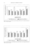
STRUCTURE AND PERMEABILITY OF HUMAN NAIL 369 was thus a measure of nail flexibility. They found that during water immersion of nails, nail weight increased by 22% of its original value in two hours and then unexpectedly decreased. Flexibility of nails continuously increased with water immersion. Treatment of nails with mineral oil showed no increase in flexibility however, mineral oil applied to previously hydrated nails prolonged their flexibility. A phospholipid-water prepara- tion was also found to increase nail flexibility of normal and lipid-extracted nails. It was postulated that this increased flexibility may be due to the ability of the phospholipids to bind water. In a more recent study, transonychial water loss (TOWL) was measured using an evaporimeter in 21 healthy adults, and was reported to be in the range of 1.17-3.35 mg/cm2/h. This rate of water loss through the nail plate was not significantly different from the transepidermal water loss (TEWL) from the dorsum of the hand. The TOWL appeared to decrease with age and was not affected by nail plate thickness (31). PERMEABILITY OF THE HUMAN NAIL By virtue of its chemical composition, the nail plate forms an effective barrier to the permeation of drugs. Thus, the diffusion of actives into and through the nail is extremely poor relative to other membranes such as the skin. Hence, topical medication for treatment of nail infections has been ineffective to date, and information on nail per- meability still remains limited. Currently, only oral antifungal therapy is approved for treatment of OM in the United States. However, since efficacious topical nail therapy is most desirable, recent research has focused on characterizing and improving the perme- ation of drugs through the human nail. Some of the permeability properties and advances made in topical therapy of nail diseases are discussed in the remainder of this review. EFFECT OF MOLECULAR WEIGHT AND LIPOPHILICITY OF THE PERMEANT Walters et al. designed the first diffusion cell, specially adapted to quantitatively mea- sure nail plate permeability in vitro, in 1981 (32). This stainless steel diffusion cell permitted the exposure of 0.38 cm 2 of the nail plate to the donor and receiver solutions, present on either side of the nail. The flux and permeability coefficients of the per- meants, based on Fick's Law, were reported. A water flux (determined by monitoring the permeation of tritiated water through cadaver nails) of 12.6 + 5.8 mg/cm2/h was obtained using this diffusion cell. This flux value was about five times higher than that reported by earlier researchers. This was explained by the fact that nails used in this study were hydrated, in contrast to dry nails used in earlier investigations. To check the integrity of the nail permeability barrier as a consequence of hydration, the permeability coefficient of methanol was monitored over a period of 49 days and was found to remain fairly constant. This indicated that the nail was a stable barrier and was not affected by the repeated application of pressure required to seal the nail in the diffusion cell. Furthermore, the nail plate permeability was found to be almost inversely proportional to nail thickness. These diffusion cells were later used to study the in vitro permeation of a series of dilute, homologous alcohols (from methanol to n-dodecanol) through the nail plate. A radio- active assay was used to analyze concentration of the active, and permeability coeffi-

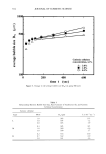
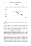
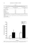
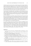
370 JOURNAL OF COSMETIC SCIENCE cients, lag times, and effective diffusion coefficients were calculated. The effect of increasing chain length on the permeability coefficients and diffusion coefficients of the n-alkanols is depicted in Figure 2. Figure 2 shows that the permeability coefficients and diffusion coefficients of the alcohols from methanol through n-octanol decreased with increasing chain length. The lag times also showed a corresponding increase (not shown). The authors thus concluded that the nail plate appeared to behave as a concentrated hydrogel in this case, and that the partitioning of the alcohols into the complex keratin matrix decreased with increasing hydrophobicity up to n-octanol. However, with the higher alkanols from n-octanol to n-dodecanol, it was found that the permeability coefficients through the nail plate increased exponentially (Figure 2). The authors sug- gested the involvement of a possible parallel lipid pathway to explain the permeability result for extremely hydrophobic molecules (33). Neat (undiluted) alcohols were also found to demonstrate trends similar in permeation to the dilute alcohols. However, the permeability coefficients of the undiluted alcohols (except methanol) were about one fifth those of their dilute counterparts. It was observed that the rate of transfer for the lower alcohols increased through the lipid-extracted (with a 3:1 chloroform:methanol mixture) nail plate. On the other hand, a sixfold decrease in the permeation rate of n-decanol was observed through the delipidized nail. The authors suggested that the decrease in permeation rate observed for the larger alkanols resulted because the rate- 18.00 6.00 16.00 14.00 12.00 10.00 8.00 6.00 4.00 2.00 0.00 A Permeability Coefficient - ß- Diffusion Coefficient ß 0---0... 0 ß 0 2 4 6 8 10 12 5.00 4.00 3.00 2.00 1.00 0.00 14 Alcohol chain length Figure 2. Effect of chain length on the permeability coefficients and diffusion coefficients of a series of diluted, homologous n-alkanols through the human nail plate. Mean _+ S.D., n = 4-26. Adapted from reference 33.
Purchased for the exclusive use of nofirst nolast (unknown)
From: SCC Media Library & Resource Center (library.scconline.org)