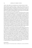
STRUCTURE AND PERMEABILITY OF HUMAN NAIL 377 efficacy of a topical antifungal treatment. This coefficient was defined as shown in equation 2: E = Jm,•.,JMIC (2) whereJ,,a:• represents maximum flux, and MIC is the minimum inhibitory concentration of the antimycotic drug. Thus, enhanced penetration (high J ..... ) of a potent antifungal (low MIC) would result in an efficacious topical formulation for treatment of OM. In an early study, the effect of petrolatum, ethylene glycol monomethyl ether (EGME), dimethylacetamide, and dimethyl sulfoxide (DMSO) on nail penetration, in ten subjects, was investigated. A fluorescent dye was used as a marker, and depth of penetration was judged by scraping the nail plate with a knife. Penetration was found to be insignificant with petrolatum and dimethylacetamide. In the case of DMSO and EGME, a maximum penetration of about one fourth the depth of the nail plate was achieved (42). In another study, the conventionally accepted skin penetration enhancer, DMSO, was shown to retard the permeation rate of methanol and hexanol across the nail plate. Isopropanol was found to reduce the rate of nail permeation of hexanol, while it had minimal effect on the permeability of methanol (43). Skin penetration enhancers, such as DMSO, are thought to interact with the lipid domains of the stratum corneum by increasing the fluidity or increasing the partitioning of the drug into it. Due to the low levels of lipid in the nail and the possibly less developed lipid pathways, skin penetration enhancers may be ineffective in accelerating nail penetration (5). Soong (35) demonstrated that treatment of the nail with agents such as dithiothreitol (containing sulfhydryl groups) resulted in enhanced permeation for various model com- pounds (acetic acid, benzoic acid, and suprofen). The effect of treatment of the nail with dithiothreitol on the lag times and diffusion coefficients of the three drugs is shown in Table IV. As seen in Table IV, for all three compounds investigated, dithiothreitol treatment resulted in a reduction in lag times and an increase in diffusion coefficients. The greatest effect was observed with the compound of highest molecular weight (su- profen). The enhanced permeation for all the model compounds was attributed to the reduction of the disulfide linkages in the keratin matrix of the nail plate by dithio- threitol and, thus, an increased diffusivity within the nail for the drug. The author suggests that such chemical treatment would be particularly useful in enhancing the permeability of large, charged molecules, which would otherwise have extremely poor nail penetration rates. Table IV Effect of Treatment of Nails With Dithiothreitol on the Lag Times (tL) and Diffusion Coefficients (D) of Various Drugs, Shown as the Ratio of the Parameters of the Treated (t) to the Untreated (u) Nails Permeation parameter of treated nail (t)/ permeation parameter of untreated nail (u) Model Molecular Lag time Diffusion coefficient compound weight tL(t)/tL(Z•) D(O/D(•) Acetic acid 60 0.62 8.3 Benzoic acid 122 0.47 11 Suprofen 260 0.11 45 Adapted from reference 35.

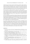
378 JOURNAL OF COSMETIC SCIENCE The influence of keratolytic agents (urea, salicylic acid, and papain) on the permeation of imidazole antimycotics (miconazole nitrate, ketoconazole, and itraconazole) through the human nail was studied in vitro (44). The experiments were carried out using side-by-side diffusion cells, and the donor and receptor solution were composed of a 60:40 ethanol:water mixture. To assay the drug content, the samples were reacted with bromocresol green, and the complex between the imidazole moiety and the dye was measured spectrophotometrically. It was reported that ethanol (used as a co-solvent) did not promote the passage of any of the antimycotics through the nail over a period of 60 days. Scanning electron microscopy studies revealed that all the keratolytic agents modified the normal scaly surface of the nails (as evidenced by a more fractured scaly surface). However, pretreatment of the nails with salicylic acid alone (20% for ten days), or application of the drug in a 40% urea solution, was not effective in enhancing nail permeation. Only the combined effect of pretreatment with papain (15% for one day) and salicylic acid (20% for ten days) was effective. The authors postulated that the keratolytic action of papain in conjunction with salicylic acid results in the formation of pores in the nail, thereby creating transport channels for the drug to permeate. Novel ways of using chemical enhancers to directly interact with sites on nail keratin were recently reported (5,45). A schematic diagram of the bonds in nail keratin, which represent potential interaction sites for nail penetration enhancers, is depicted in Figure 5. In the patented innovation of Sun eta/. (45), N-acetyl-l-cysteine (AC) and urea were S Disulfide Linkage S S T Keratin Protein Chai Peptide N Linkage [ COO-- Polar Linkage c H Hydrogen Linkage Figure 5. Schematic representation of the bonds in nail keratin that represent potential interaction sites for nail penetration enhancers. Adapted from reference 5.
Purchased for the exclusive use of nofirst nolast (unknown)
From: SCC Media Library & Resource Center (library.scconline.org)