118 JOURNAL OF COSMETIC SCIENCE EFFECT OF DEOXYARBUTIN ON MELANOGENESIS: IN VIVO COMPARISION WITH OTHER MELANOGENESIS INHIBITOR Saja H. Hamed, Penkanok Sriwiiyanont, R. Randall Wickett, Ph.D. and Raymond Boissy, Ph.D. University �l Cincinnati College of Pharmacy and Department �/Dermatology, Cincinnati, Ohio Introduction: Skin color is mainly determined by the amount of melanin in the epidermis. Melanin biosynthesis occurs in specialized organelles called melanosomes, which are synthesized within the melanocytes dendritic cells of neural crest origin that resides in the basal layer of the epidermis ( 1 ). Through dendritic processes, melanocytes transfer pigmented melanosomes into neighboring keratinocytes. Tyrosinase, an enzyme present within melanosomes catalyses the initial step in the conversion of tyrosine to melanin (1). Competitive inhibitors of tyrosinase (hydroquinone, kojic acid, and arbutin) are currently being used in drugs to ameliorate hyperpigmented lesions and in cosmetics to lighten skin complexion. Although these skin lightening products have been available for several decades, their success has been somewhat limited for two basic reasons: safety or overall effectiveness, Therefore, to create a better and non-irritating tyrosinase inhibitor, a rational design approach was used that evaluated structure -function relationships of tyrosinase inhibitors. One designed compound, deoxyarbutin ( dA), demonstrated effective skin depigrnenting properties using a black guinea pig model. The research project in this report seeks to ascertain the effectiveness of deoxyarbutin in inhibiting melanin synthesis within human melanocytes and its effectiveness in inhibiting hyperpigrnentation of human skin grafted to athymic mice. Human cutaneous xenografts placed on athymic mice survive permanently (2). Thus athymic animals are an excellent model for studying various aspects of human skin including hyperpigrnentation of grafted skin on burn patients. Methodology: Xenografting: Female ICR-SCID (Taconic, NY) kept under pathogen-free condition (Children's Hospital Research Foundation, Cincinnati, OH) were shaved with an electric clipper to remove the dorsal hair. The mice were anesthetized by isofluorane/oxygen (3o/cJ0.8 liter). The dorsal site was cut to produce a wound bed of approximately 2.0-3.0 in diameter. Fresh split-thickness cadaveric skin (Ohio Valley Tissue and Skin Center, OH) from a caucasian donor was sutured in place with a reversed cutting precision monofilament PS-3, 6-0 (Moore medical, CT). Grafts were left untreated for two months during which time hyperpigrnentation occurred. Stability of subsequent hyperpigmentation was assessed on weekly intervals using Charrnview system. Treatment was initiated when no further increase in pigmentation was observed. Animals were randomized according to their L value among four treatment groups (deoxyarbutin (dA), hydroquinone (HQ), 4-tertry butyl phenol ( 4-TBP) and control group). Each group had 4 mice. Treatment was applied at 5% concentration 5 days per week for 8 weeks. Treatment sites were assessed on biweekly basis for degree of pigmentation using Charrnview system. This system takes enlarged digital photos for the treatment sites and then the color parameters for these images (L, a, b) were obtained by using Photoshop software (Adobe Systems Inc., San Jose, Calif.). The L * a* b system is recommended by the CIE (Commission Internationale de I'Eclairage) for skin color assessment. In this project the L value which represents the Luminance was used to assess the lightening effect of the applied treatments. Cytotoxicity: Cultures of normal human melanocytes was established from individual neonatal foreskins that were obtained from the nursery of University Hospital after routine circumcision. Established melanocytes from a light and a dark skin donor were treated with fresh growth media containing various dosages of test compounds for 5 days. On the 6th day, cell number was determined by direct counting using the Coulter Counter. In Situ (Intact) Tyrosine Hydroxylase Assay: Cultured human melanocytes were treated in triplicate with fresh growth media containing various dosages of test compounds for 5 days. On the 5th day cells were fed with fresh media containing I µCi/ml of H3-tyrosine (Arnersham Pharmacia Biotech, Piscataway, NJ). Radioactivity of the tritium water in the elute was counted in a Packard 1600 CA liquid scintillation analyzer. Tyrosinase activity was expressed as DPM/24 hours/µg protein.

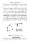
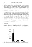
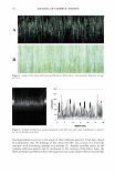
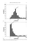
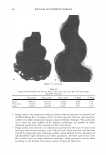
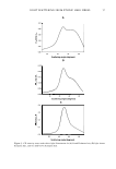
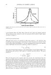
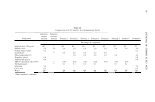
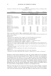
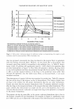
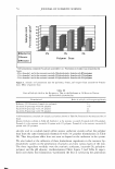
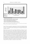
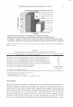
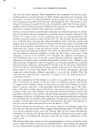
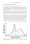
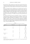
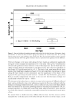
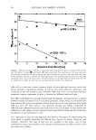
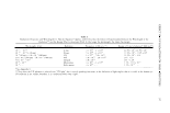
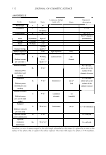
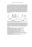
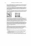
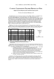
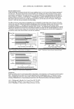
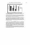
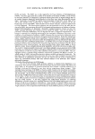
2003 ANNUAL SCIENTIFIC MEETING Results: Deoxyarlmtin was less cytotoxic than hydroquinone by 4 fold on both light and dark human melanocytes (Table 1). Further more deoxyarbutin was as effective as hydroquinone in inhibiting In situ tyrosinase activity of dark human melanocytes at equimolar concentrations (Figure 2). Table I. Concentration (µM) of each test compound that lead to 95% viability of dark and light human melan ocvtes. Compound Dark human melanocytes Light human melanocytes hvdroauinone 0.391 1.5625 deoxvarbutin 1.5625 6.25 4- tertrv butvl phenol 50 50 koiic acid 50 50 arbutin 50 50 Figure 2.Effect of 5 days of treatment with deoxyarbutin and hydroquinone f 1.2 ,_,,__,,_�- -- ··•···dAUeatedcell (Ty..-oalnaae catalytic actlvlty(DPM/1'!1 prtn per control)) on In situ tyrosinase activity of dark human melanocytes. " I 0.6 +--�-�--=-- - i ! 0.4 +- - - - - - - - - 1 0.2 +- - - - - - - - - -- --a--- HQ treated cell (Ty.-oelnaae catalytic actlvlty(DPM/1-19 prtn rcontrol ! 10 Cone (mlcromol■r) 15 Animal study showed that both deoxyarbutin and hydroquinone are able to reverse graft hyperpigmentation (Figure 3). However the mean value for Change in L from the baseline was higher for deoxyarbutin treated group at the 2 weeks treatment point compared to hydroquinone. Brown discoloration of the hair in hydroquinone treated mice was observed. Such brown discoloration was not observed in the deoxyarbutin treated mice. 30.00 25.00 20.00 I 15.00 � 10.00 5.00 0.00 2wks-d0 Change In L value from the ba-llne 4wks-d0 Swks-d0 8wks-d0 ---+- Cont ···•·· HQ --4--dA TSP Figure 3.Time course of change in L value from treatment with Deoxyarbutin, Hydroquinone and 4-TBP Conclusion: The ability of deoxyarbutin to inhibit pigmentation with reduced cytotoxicity relative to hydroquinone establishes deoxyarbutin as an excellent skin depigmentation agent. References: 1. Boissy RE and Nordlund JJ: Biology of Melanocytes, In: Arndt KA, LeBoit PE, Robinson JK, Wintroub BU ( eds): Cutaneous Medicine and Surgery: An Integrated Program in Dermatology. Philadelphia, WB Saunders Co, pp. 1203-1209 (1995) 2. Manning DD, Reed ND, Schaffer CF: Maintenance of skin xenografts of widely divergent phylogenetic origin of congenitally athyrnic (nude) mice. J Exp Med 133: 488-494 (1973) 3. U.S patent no. 6,068,834 (5/30/00) 119
Purchased for the exclusive use of nofirst nolast (unknown)
From: SCC Media Library & Resource Center (library.scconline.org)