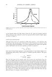
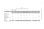
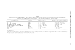
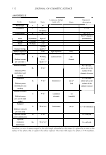
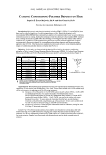
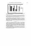
132 JOURNAL OF COSMETIC SCIENCE BASICS OF SKIN STRUCTURE R. Randall Wickett, Ph.D. University of Cincinnati College of Pharmacy, Cincinnati, Ohio Introduction to the skin The skin is the largest organ of the human body accounting for about 16% of total body weight. Its vital role Is to keep us in and the world out. The skin also has immune functions and helps to regulate body temperature. Understanding the skin especially the Sratum Corneum is important to the cosmetic scientists as cosmetic products are bound to come in contact with the skin. Layers of the skin The main structural components of the skin are the dennis, the epidennis. The dermis is divided into two layers, the papill.ary dermis and the reticular dermis. The dermis provides the bulk of the mechanical strength to the skin. The hypodermis which contains the subcutaneous fat is beneath the dermis. Growing hair follicles are rooted in the hypodermis. The epidermis is subdivided into five strata, basal, spinous, granular, lucid and corneum. The top layer, the Stratum Corneum (SC) is the primary barrier to transport of water and other molecules across the skin. This lecture will focus on the formation and structure of the Stratum Corneum. Nomenclature for I avers of the Eoidermis Eni:ilish Latin Alternative Basal cell laver Stratum Basa/e S. Germinativium MalphQhian layer Prickle Laver S. Spinosum Maloiahian laver Granular Laver S. Granulosum Maloiohian laver Clear Laver S. Lucidum Horny Layer S. Corneum The major cell type of the epidermis is the keratinocyte which makes keratin proteins. Keratins are fibrous proteins of the epidermis that are the major structural proteins of SC hair and nail. Keratins are in class of proteins called intermediate filaments which from part of the cytoskeleton of all nucleated cells. Formation of the SC barrier The epidermis continually renews. Slow cycling stem cells at the basal layer divide and one daughter cell remains as a slow cycling stem cell and the other becomes a transient amplifying cell. T.A. cells continue to divide until they become post mitotic and terminally differentiate. Specific keratins (k1 and k10) are expressed as markers of the transition from proliferative TA cells to terminally differentiated keratinocytes that will be committed to forming SC. At the Stratum Granu/osum(SG) keratohyalin granules full of protein and lamellar bodies appear in the cells. Then the cells are transformed to squames. The nucleus is digested, the cytoplasm disappears, lipids are dumped into the intercellular space, the keratin filaments aggregate to microfibrils and the cell membranes is replaced by a cell envelope. As a result of transformations at the SG the SC barrier is formed. 15 - 20 layers of cells Flattened cells (corneocytes or squames) with resistant cell envelope and attached lipids Content is keratin microfibrils Squames are joined by desmosomes. lntercellular lipids Multiple layers between cells, polar but relatively hydrophobic Ceramides, cholesterol, cholesterol esters and long chain fatty acids

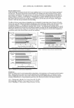
2003 ANNUAL SCIENTIFIC MEETING The Bricks and Mortar Model The permeability barrier of the SC is sometimes modeled as a brick wall with the bricks being the corneocytes with the resistant cell envelopes and keratin microfibrils and the mortar being the intercellular lipids. The mortar is the main barrier to water passing through the SC and lipid soluble molecules are modeled as winding their way through the mortar. The bricks (corneocytes) are 70-80% keratin by dry weight. The keratin is condensed in the form of microfibrils. The keratinocyte cell membrane is replaced by a tough new structure of cross linked protein called the cell envelope. There are lipids covalently attached to the cell envelope on the outer surface and the corneocytes are joined by desmosomes. The keratins in keratinocytes below the SG are in the form of coiled-coiled coils of ex-helix. As the SG cells are transformed into squames the coiled coils aggregate to form 32 chain structures called microfibrils which lay parallel to the surface of the skin and restrict in plane swelling of the squames. Two proteins from the keratohyalin granules, filaggrin and loricrin play key roles in the fonnation of the "bricks". Filaggrin is an acronym for filament aggregating protein. Filaggrin contain a high level of positively charge amino acids and participates in the aggregation of the negatively charged keratin coiled coils. Loricrin is a globular protein that is rich in hydrophobic amino acids and cysteine. It is released from the granules and is crossed-linked to the protein involucrin which is already in the cytoplasm of the cell. The two proteins are cross-linked by the membrane bound enzyme transglutaminase to begin forming the cell envelope. The crosslink is formed between lysine and glutamic acid side chains to form what is known as the isopeptide bond. Eventually the entire cell membrane is replaced by cross-linked protein and lipids (ceramides) are covalently attached to the outer surface of the this new structure which is known as the resistant cell envelope. Keratin microbrils on the inside are also cross-linked to the envelope. The lamellar bodies that appear at the SG contain lipids which are released into the intercellular space as the SC forms. These lipids are glucosyl ceramides, cholesterol, cholesterol esters and long chain fatty acids. In the intercellular space the glucosyl cermides are converted to ceramides and phosopholipids from the original cell membrane are degraded to fatty acids. The SC contains no phospholipids. The lipids in the intercellular spaces arrange themselves into multiple layers. These multilamellar lipids are the mortar of the bricks and mortar model of SC barrier function. References: Marekov and Steinert, Ceramides are bound to structural proteins of the human foreskin epidermal cornified cell envelope, J. Biol Chem. 273, 17763-17770 (1998) Steinert, P.M., Marekov, L.N., Fraser, R.D.B. and Parry, D.A.D. Keratin intermediate filament structure: Crosslinking studies yield quantitative information on molecular dimensions and mechanics of assembly. J Mo/ Bio 230, 436-452. (1993) Steinert and Marekov, Direct evidence that involucrin is a major early isopeptide cross-linked component of the keratinocyte cornified cell envelope. J. Biol Chem. 272:2021-2030(1997) Swartzendruber, D. C., Wertz, P. W., Kitko, D. J., Madison, K. C., & Downing, D. T Molecular models of the intercellular lipid lamellae in mammalian stratum corneum. J Invest Dermatol, 92(2): 251-257. (1989.). Wertz, P. W. & Downing, D. T. Covalently bound omega-hydroxyacylsphingosine in the stratum corneum. Biochim Biophys Acta, 917(1): 108-111. (1987). 133
Purchased for the exclusive use of nofirst nolast (unknown)
From: SCC Media Library & Resource Center (library.scconline.org)