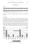
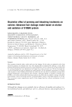
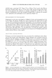
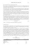
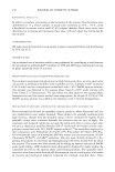
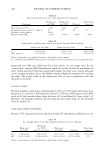
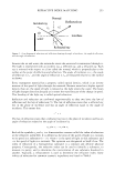
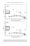
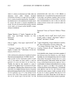
222 JOURNAL OF COSMETIC SCIENCE 2D GEL-ELECTROPHORESIS OF SlO0A3 IN DELAMINATED CUTICLES Delaminated cuticles (1 mg) from bleached hair were extracted with 100 mM dithio threitol containing 200 mM Tris-HCl buffer (pH 7 .6). Extracts were concentrated by precipitation with trichloroacetic acid. The precipitate was applied to modified two dimensional electrophoresis, as previously described (3 ). Isoelectric focusing was per formed according to standard protocols using IPG strips with a narrow pH gradient of 3-5 (Sigma-Aldrich, St. Louis, MO). The second dimension was performed under the same conditions described above, using the Zoom type of Nu-PAGE gel at 7 .8 x 6.3 x 0.1 cm. The mean density of spots and their proximal background zone on silver-stained 2D-P AGE gels were measured using image-analyzing software (Scion). Subtracted mean densities were used for proportion calculation. RESULTS At first, we examined whether the turbidity (OD 600 ) accurately represents the concen tration of the cuticles in suspension. OD 600 was linearly increased up to 1.0, depending on the amount of suspended cuticle (Figure la). No difference in OD 600 was observed among non-treated, permed, and bleached hair samples. Consistent with previous re ports (7), the cuticles of both permed and bleached hair were more easily delaminated compared to untreated ones (Figure 16,c). The amount of delaminated cuticles from bleached hair was increased as a higher concentration of hydrogen peroxide was applied. In the case of permed hair, however, application of an excessive concentration (3%) of thioglycolate results in reduction of the delamination. This might be attributed to the lesser rubbing of the wavy hair with the water-stirring method. Due to their very flat shape and their transparent nature, it is difficult to clearly identify delaminated cuticle fragments by conventional microscopy. In this study, we observed the autofluorescence of delaminated cuticles using confocal fluorescent microscopy (Fig ure 2a). Confocal images were processed using gradation analysis software. The trans formed gradation image revealed that the average area of each cuticle fragment, delami nated from permed hair, was about twice as large as that of untreated hair (Figure 26), whereas those of bleached hair were 30% smaller compared to the normal ones (Figure 2c). These results indicate that the physiological characteristics of cuticles from the bleached hair are distinct from those of the permed hair. We previously detected SlO0A3 in the permanent waving lotion (5). We examined the correlation of Sl00A3 elution by perming treatment with the enlargement of cuticle delamination. Treatment of hair fiber at the minimum thioglycolate concentration required to elute SlO0A3 into permanent waving lotion (�0.3%, Figure 3) resulted in significant enlargement of the cuticles. Although application of higher thioglycolate concentration increased the amount of eluted S 1 00A3 protein, the enlargement of delaminated cuticles reached a plateau at 1 % concentration (Figure 26). These results indicate that the loss of S 1 00A3 protein, even in a low amount, results in cuticle delamination. In contrast to permanent waving, bleaching did not result in a release of the Sl00A3 protein (data not shown). Nevertheless, cuticles of bleached hair were easily delaminated (Figure le) and fragmented into small pieces (Figure 2c). In this study, we performed 2D PAGE analysis of extracts of the cuticles delaminated from bleached hair. Although the

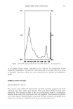
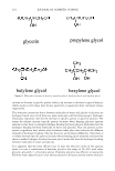
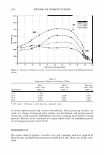
EFFECT OF PERMING/BLEACHING ON CUTICLES 223 a b 14 C 8 Perm )IOI( )IOI( Bleach 12 10 ,,...., * * ........ i i -d" 8 -d" � � 4 ( 6 ( 4 2 2 0 0 0.3 1 3 0 0 1 3 10 Thio&lyct-olate (%) �� (%) Figure 2. Effects of perming and bleaching on the size of delaminated cuticles. (a) Confocal microscopy of cuticles delaminated from permed and bleached hair. Cuticle suspensions were prepared from hair samples treated with various concentrations of thioglycolate and hydrogen peroxide (bottom row). Note the size of delaminated cuticles from permed (b) and bleached hair (c). The area of cuticle fragments was computed by gradational analysis of each confocal image. Values represent the average of three independent experiments ± SD. Statistical analyses were performed by t-test of the values obtained without hair treatment chemicals. * p 0.05 ** p 0.01. precise identification of each isoelectric variant of S 1 00A3 required further investiga tion, observed acidic shifts of the protein spots suggested that several cysteine residues in the S 100A3 protein were converted into cysteic acid by oxidation with hydrogen peroxide. Our 2D PAGE analyses showed that S100A3 with a higher content of cysteic acid occupied a higher proportion in bleached hair treated with a higher concentration of hydrogen peroxide (Figure 4). DISCUSSION Various techniques, such as morphological observation of hair fiber using scanning electron microscopy and tensile measurement, have been employed to monitor hair damage. However, the critical differences between the perm and bleach inductive dam aging mechanism(s) have never been studied. In this study, we report that the manner
Purchased for the exclusive use of nofirst nolast (unknown)
From: SCC Media Library & Resource Center (library.scconline.org)