220 JOURNAL OF COSMETIC SCIENCE hairdressing products (i.e., chemical factors) results in sudden and severe changes. Per manent waving and bleaching treatments during hair coloring processes are major causes of hair damage. In order to improve hair care products, it is essential to understand the mechanism of hair damage induced by these procedures. The scale-like cuticle surrounds the hair cortex. Normally, cuticles are damaged prior to changes occurring within the cortex. We previously identified and characterized SlO0A3, a unique member with high cysteine content of the largest calcium-binding SlO0 protein family, characterized by tissue-specific distribution (1), in cuticles (2,3). Its soluble nature under reducing conditions is different from that of other hair proteinous components such as hair keratins and keratin-associating proteins. Ultrastructural lo calization of SlO0A3 in the inner part of cuticles (i.e., endocuticles) suggested its structural role in preserving the attachment of adjacent cuticles (4). The release of S 1 00A3 protein from the attachment site is suggested to be the most relevant event leading to hair damage. We previously proposed a hair damage model that implies the involvement of Sl00A3 in hair damaging processes (5). This model was based on the following four stages: (a) Newly emerging hair is characterized by a smooth-edged cuticle. (6) As a result of cracking of the edge of intact cuticles by normal grooming, the SlO0A3-rich layer in turn becomes the outermost layer. (c) SlO0A3, a soluble protein under non-reducing condition, is oxidized due to the cleavage of inter molecular disulfide bridges under environmental stresses such as UV radiation. (d) Sl00A3 is gradually released from hair during daily washing. It seems also possible that application of permanent waving lotion is able to elute SlO0A3 from natural hair within the second stage. However, it is still unknown how bleaching treatment induces hair damage without elution of SlO0A3. In this study, we examined how chemical hair treatments affected cuticle delamination. Confocal microscopy revealed that permanent waving and bleaching treatments had dissimilar effects on the size of delaminated cuticles. In addition, 2-dimensional elec trophoresis revealed an acidic shift of S 1 00A3 due to irreversible modification of several residues out of the total of ten cysteines in oxidized hair. Based on these results, we present a comprehensive model of hair damage induced by chemical treatments. MATERIALS AND METHODS PERMANENT WAVING AND BLEACHING TREATMENTS Hair was bleached by immersion in 1-10% hydrogen peroxide-ammonia solution (pH 9. 5) for 1 hr at 3 7 ° C. Permanent waving treatment was done by immersing hair at 37 ° C in 0.1-3% ammonium thioglycolate solution containing 1% 2-aminoethanol and 0.45% ammonia for 1 hr, followed by neutralization by 7% sodium bromate for 15 min. Hair samples were collected from males of Japanese descent. DELAMINATION OF CUTICLES The mechanical isolation method for cuticles consisting of stirring hair in water ( 6) was modified with a small-scale delamination test as follows. One-cm hair fibers (200 mg) were stirred at 180 rpm using an R30 shaker (Taitec, Tokyo, Japan) with 10 ml of

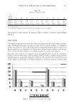
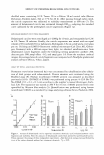
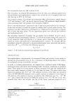
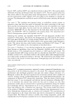
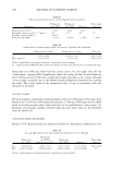
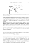
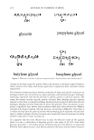
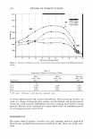
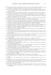
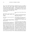
EFFECT OF PERMING/BLEACHING ON CUTICLES 221 distilled water contammg 0.1 % Tween 20 in a Falcon 50-ml conical tube (Becton Dickinson, Franklin Lakes, NJ) at 3 7 ° C for 20 hr. After passing through nylon mesh, the cuticle suspension was subjected to turbidity measurement at 600 nm (7). The amount of delaminated cuticle was estimated through OD600, adopting the standard curve calibrated by the preweighed cuticle suspensions (Figure la). SIZE MEASUREMENT OF CUTICLE FRAGMENTS Delaminated cuticles were centrifuged at 14,000g for 10 min, and resuspended at 0.2% in 1 % Tween 20 solution. Finally, the cuticle suspension was mixed with an equal volume ofVectorshield (Vector Laboratory, Burlingame, CA) on a slide and a cover glass put on. Utilizing an LSM510 fluorescent confocal microscope (Carl Zeiss AG, Gottin gen, Germany) with a 488-nm argon laser light, we observed autofluorescence from delaminated cuticle fragments under the following setting parameters: pinhole: 200 detector gain: 900 ampl offset: -0.2 and ampl gain: 1.0. From the resultant confocal images, the averaged area of cuticle fragments was computed with NewQube gradational analysis software (Nexus, Tokyo, Japan). ASSAY OF TOTAL AND Sl00A3 PROTEINS Permanent-wave-lotion-immersed hair was concentrated by ultrafiltration after alkyla tion of thiol groups with iodoacetamide. Protein amounts were estimated using the Bradford assay (8). Human recombinant S100A3 protein was prepared as described previously (9,10). ID-PAGE of S100A3 was performed using precast 4-12% Bis-Tris Nu-PAGE (7 .8 x 6.3 x 0.1 cm) according to the supplier's manual (Invitrogen, Carls bad, CA). The separation was started at 200 V for 3 5 min. S 1 00A3 protein levels were quantified by Western blot analysis (5 ). Quantification was performed, using human recombinant S100A3 as a standard, by image-analyzing software (Scion, Frederick, MD). a b C 1.0 � 0. 7 � 0. 9 Bleach * Perm * "' )OB J 0.6 0.8 I.. � 0.7 * i 0.5 (U ' � 0.6 0 0.6 � 0.4 � 0.5 □� · · D 0.4 • l\bn-treat a 03 a 0.4 •Perm ""Cl "i 0.3 (1) 1ii 0.2 +-' 0.2 A Bleach .£ -� 0.2 E 0.1 j 0.1 J!! (1) (1) 0 0 0 0 0.010 0.020 0 0.3 1.0 3.0 0 3 10 Cuticle (w/� Thioglych:llate (%) f-i:i02 (%) Figure 1. Increase of delaminated cuticles upon hair treatment chemicals. (a) Correlation between cuticle fragments and turbidity. Note the amounts of delaminated cuticle from permed (b) and bleached hair (c). Values represent the average of three independent experiments ± SD. Statistical analyses were performed by t-test of the values obtained without hair treatment chemicals. * p 0.05.
Purchased for the exclusive use of nofirst nolast (unknown)
From: SCC Media Library & Resource Center (library.scconline.org)