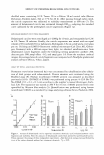
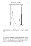
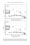
232 JOURNAL OF COSMETIC SCIENCE potassium iodide, and for this reason the peroxide number can be determined by io dometric titration. The peroxide number is the number of active oxygen milliequiva lents present in 1000 g of fat mass, which correspond to the milliequivalents of iodine released from potassium iodide titrated with sodium thiosulphate solution. Degraded oils may have peroxide numbers well below expectations, since the first step of lipoperoxidation, auto-oxidation, may already be completed, and all the hydroperox ides derived from this step may have been transformed into secondary volatile unpleas ant-smelling compounds. The peroxide number was determined as follows: Five grams (m2) of oil was placed in a 250-ml Erlenmeyer flask, and the flask was closed with an emery cap. Thirty milliliters of chloroform/acetic acid (2:3) mixture was then added to the oil sample under stirring. After complete dissolution, 0.5 ml of potassium iodide saturated solution (R2) was added. The solution was stirred for one minute, and 30 ml of distilled water was added. The mixture was then titrated with 0.01 M sodium thiosulphate solution, added slowly under continuous stirring until the yellow color disappeared. Five milliliters of starch indicator was then added and a dark blue color appeared. The titration continued during addition of sodium thiosulphate solution and vigorous stirring until the blue color disappeared (n2 ml of 0.0lM sodium thiosulphate solution). A control titration was carried out on a reference sample under the same conditions. The final volume (n 3 ml of 0.0lM sodium thiosulphate solution) employed for the control titration must be less than 0.1 ml. The peroxide number, expressed in milliequivalents of oxygen per 1000 grams of fat, is calculated from the following equation: 10(n2 - n 3 ) Peroxide number = --- - m2 DETERMINATION OF CONJUGATED DIENES AND TRIENES (5) Spectrophotometric examination can provide information on the quality of a fat, its state of conservation, and any changes produced in it by technological processes. Absorption at the wavelength 232 nm is due to the presence of conjugated diene systems, while trienes absorb at 262, 268, and 274 nm. The normal spectrum of a non-rancid virgin oil shows no absorption at these wavelengths. Oil was dissolved in 1-butanol, spectropho tometrically pure in the range of wavelengths considered. Extinctions at the various wavelengths were then detected with reference to pure solvent. The absorbence values were expressed as specific extinctions E 1 % 1cm ( the extinction of 1 % solution of the fat in the specified solvent, at a thickness of 1 cm), conventionally indicated by K, also referred to as the "extinction coefficient." In accordance with the official method in the EEC regulations, spectrophotometric analysis of oil involves determining the specific extinction at a wavelength of 232 nm and determining the variation in specific extinc tion, which is given by the following equation: (6)

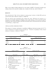
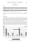
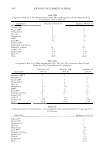
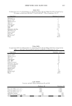
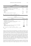
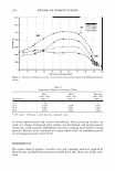
HEMP-SEED AND OLIVE OILS 233 For non-rancid virgin oils, D..K is below 0.010. Pure oil analysis. To simulate the degradation of oil over time, an accelerated stability test was performed: spectrophotometric analysis was carried out on oil samples before and after heating to 40°C for 24 hours. Oil-in-emulsion analysis. To evaluate the antioxidant effect of excipients, namely Sepigel 305® and Carbopol 1382®, on the oil dispersed in an 0/W emulsion, simple formula tions containing these two products were prepared, (Table I). Prior to spectrophotometric examination the oil had to be extracted from the emulsion. It was thus necessary to break the system by adding NaCl crystals under stirring. The dispersion was then subjected to centrifugation at 13000 rpm for 10 minutes to separate the oil from the water phase. The fat supernatant phase was collected and analyzed spectrophotometricall y. The procedure employed to prepare the oil samples was as follows: A 0.1 % oil-in- 1-butanol solution was prepared, placing 27 µl of oil (oil density being 0.92 g/ml) in a 25-ml volumetric flask and diluting it to volume with 1-butanol. The solution was stirred on vortex and subjected to spectrophotometric analysis. To calculate D..K the absorbance values (E,.) detected by the spectrophotometer were converted into specific extinctions (E 1 % lcm or K) from the following equation: KA= EA.IC (7) where C is the solution concentration expressed in g/100 ml. IRRADIATION TEST Spectrophotometric determination of conjugated dienes and trienes is also useful to evaluate the photostability of an oil. As a consequence of photodegradation, the number of conjugated systems considerably increases. In order to evaluate photostability, oil samples were irradiated under UVB lamps. Conjugated dienes and trienes were determined after two hours' irradiation, as described above. Three grams of oil were placed in Pyrex containers with caps and irradiated for two hours under magnetic stirring, at 10 cm from the lamp. The mean surface area exposed to radiation was 10.17 cm2. The radiation intensity of the lamp was measured with a CO.FO.ME.GRA measurer with a probe sensitive to radiation in the range of 290-400 nm. The radiation intensity recorded for the UVB lamp was 2.6 x 10⁻⁴ W cm- 2 this is double the intensity ofUVB solar radiation (1.3 x 10⁻⁴ W cm- 2 ) (12). Two variables were taken into account: the cap on the Pyrex containers might act as a Ingredients Vegetable (hemp-seed, olive) oil Sepigel 305 ® Carbopol 1382® NaOH 10% in water Water Table I Composition of Formulations Emulsion 1 (w/w %) 5 94 Emulsion 2 (w/w %) 5 0.25 0.27 94.98
Purchased for the exclusive use of nofirst nolast (unknown)
From: SCC Media Library & Resource Center (library.scconline.org)