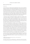
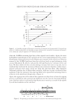
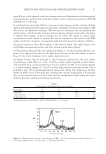
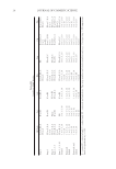
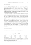
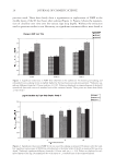
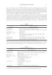
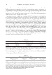
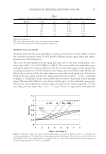
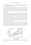
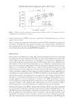
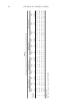
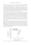
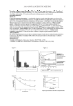
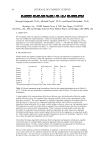
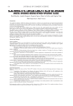
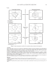
REDUCTION-INDUCED HAIR SURFACE MODIFICATION 11 outer β-layer of the exposed scale faces during reduction (delipidation) and subsequently during treatment with the low-molecular-weight cationic conditioning molecule CETAB (relipidation or refatting). X-ray photoelectron spectroscopy (XPS) as a measure of surface damage caused by reduction. To help explain and understand the changes in the surface chemistry of reduced hair samples, XPS was used as an additional technique. XPS was used to determine the concentration and chemical state of all detectable elements. In investigating changes in the surface chemistry of reduced hair samples, of special interest was, of course, the change in surface sulfur concentrations from sample to sample. For ease of comparison, the results of the XPS analysis of the hair categories investigated (unaltered and reduced) are shown in Table I. Of special interest in these XPS analyses is the element sulfur. The sulfur listed is the total sulfur concentration on the scale faces of each specifi c hair category: (a) The untreated hair had the least amount of sulfur (~0.3 atom%) among the hair cate- gories. It is expected that most of the lipid layer (F-layer) on the hair surface is intact however, low levels of damage to the surface lipids pre-exist. (b) Figure 8 shows that for both the 5- and 30-minute reduced hair, the scale surface concentration of SOx - 3 (SO ) at ~168 - 169 eV is rather small (compared to photochemi- cally oxidized hair), arising mainly from the air-oxidation of -SH. A tiny bump appears at a lower binding energy of ~164 eV on both light-exposed and reduced hair, and is probably a trace of C-S stemming from the amino acid cystine. For comparison, we have shown an XPS scan of UV-treated hair, showing the extreme delipidation UV radiation does to the exposed scale faces. It is clear that the mechanism of lipid removal is quite different between oxidative and reductive processes. Figure 8. High-resolution spectra of the changes in - x SO and C-S concentrations on the surface of (-) 200-h light-exposed, and (-) 5-min- and (…) 30-min-reduced hair. Table 1 Concentration† of Elements Detected (in atom%) Hair sample O N C S Unaltered 10.0 1.5 83.9 0.3 5-min reduced 14.3 0.8 77.6 0.6 30-min reduced 15.0 1.1 77.2 0.4 † Concentrations are normalized to 100%. XPS detection is ~0.1 atom%.

JOURNAL OF COSMETIC SCIENCE 12 CONCLUSIONS Microfl uorometric scanning, single-fi ber wettability scanning, and XPS analysis show rather similar results, in spite of their greatly varied applications. These techniques are highly sensitive to measuring changes in the surface chemistry of the scale faces. Short- term, rapid adsorption of the cationic fl uorochrome and changed wetting properties are good indicators of changes in the chemical nature and surface wettability. Since XPS analysis is able to detect atomic species at the very surface of the scale faces, receiving signals from an escape depth as shallow as 25 Å, it appeared ideal to use this technique to characterize treatment-induced changes of the hair surface. Assuming that the upper β-layer is ~50 Å-thick, XPS detects the 18-MEA domains on the scale faces prior to oxi- dation or reduction, and the newly formed sulfur entities after removal of the fatty acid 18-MEA from the scale faces. The results of these analyses clearly indicate reduction- induced delipidation and acidifi cation of the scale faces. The reduced hair fi bers show only very low concentrations of sulfur present as −SH. Sulfur present in the mixed disulfi de is not registered because of its blockage with the −CH2−COOH group. ACKNOWLEDGMENTS This work was carried out with support from TRI sponsors of the international hair-care industry. The authors thank Ms. C. J. Dansizer for wettability measurements. REFERENCES (1) S. B. Ruetsch and Y. K. Kamath, Change in surface chemistry of the cuticle of human hair by chemical and photochemical oxidation, IFSCC Magazine, 7(4) (October–December 2004). (2) S. B. Ruetsch, Y. K. Kamath, and H.-D. Weigmann, Photodegradation of human hair: An SEM study, J. Cosmet. Sci., 51, 103–125 (2000). (3) J. D. Leeder and J. A. Rippon, Changes induced in the properties of wool by specifi c epicuticle modifi - cation, J. Soc. Dyers Colour., 101, 11–16 (1985). (4) D. J. Evans, J. D. Leeder, J. A. Rippon, and D. E. Rivett, Separation and analysis of the surface lipids of wool fi ber, Proc. 7th Int. Wool Text. Res. Conf., Tokyo, 1, 135–142 (1985). (5) U. Kalkbrenner, H. Korner, H. Hocker, and D. E. Rivett, Studies on the lipids of the wool cuticle, Proc. 8th Int. Wool Text. Res. Conf., Christchurch, 1, 398–407 (1990). (6) A. P. Negri, H. J. Cornell, and D. E. Rivett, Effects of processing on the bound and free fatty acid levels in wool, Textile Res. J., 62, 381–387 (1992). (7) P. W. Wertz and D. T. Downing, Integral lipids of human hair, Lipids, 23, 878–881 (1988). (8) Y. K. Kamath, C. J. Dansizer, and H.-D. Weigmann, Wetting behavior of human hair fi bers, J. Appl. Poly. Sci., 22, 2295–2306, (1978). (9) S. B. Ruetsch and Y. K. Kamath, Fluorescence and scanning electron microscopic characterization of cuticle erosion in human hair, IFSCC Magazine, 9(1) ( January/February 2006).
Purchased for the exclusive use of nofirst nolast (unknown)
From: SCC Media Library & Resource Center (library.scconline.org)