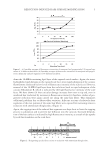
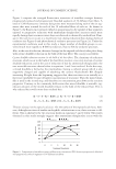
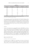
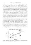
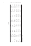
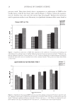
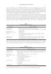
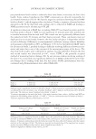
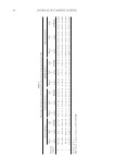
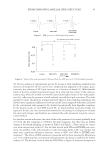
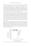
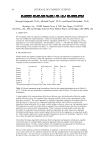
EFFECTS OF LIPID EXTRACTION AND SOAKING 21 INSTRUMENTAL RESULTS Extraction of normal forearm skin for ten minutes with 1:1 acetone/ether did not signifi - cantly change TEWL or MAT (Tables IV and V) relative to the initial skin condition on the measurement sites. Exposure of the sites to a fresh-water soak alone did not cause signifi cant changes in TEWL over the four-hour duration of the study, but did cause a signifi cant decrease in MAT on the soaked site for the fi rst 30 minutes post-soaking and on the soaked and extracted site for the duration of the study. In fact, TEWL showed no signifi cant changes with treatment or time that could not be simply explained by contin- ued water evaporation from the soaked arm, indicating that neither extracting the skin nor soaking it caused signifi cant barrier damage. It is clear, then, that although no clinical barrier damage is done by either brief water soaking or by acetone/ether extraction, signifi cant changes in the NMF result from these treatments. The combination of these treatments causes particularly great NMF loss, which is important, as we speculate that soaking and extraction mimic condi- tions caused by bathing with soap. Soap and surfactant use cause a loss of lipid in the SC not caused by water use, although water alone is suffi cient to cause dryness (26). Soap use has been shown to disrupt the lipid lamellae of the SC (27), as does A/E ex- traction. The present study quantifi es NMF levels in the upper stratum corneum and examines the relationship between NMF levels and biophysical measures of barrier integrity (TEWL) and moisture accumulation. It confi rms the conclusions previously drawn (10), that a simple ten-minute fresh-water soak is capable of removing signifi cant amounts of the free amino acid components of the stratum corneum. It also is consistent with our conclusion that SC recovery to its baseline condition after soaking is relatively slow. We also present a relatively simple, quantitative method for the analysis of the amino acid components of the NMF that discriminates the effects of common SC treatments. We believe this method will have important use for the evaluation and formulation of skin care products. SUPPLEMENTARY INFORMATION The complete, numeric NMF data sets are available by correspondence. Table IV Transepidermal Water Loss (TEWL) Study 1 Baseline .25 Hours post-soak .5 Hours post-soak 4 Hours post-soaka Control 6.3 ± 1.1 6.2 ± 0.3 6.3 ± 0.2 6.7 ± 0.2 Extracted 6.5 ± 1.9 6.5 ± 0.3 6.5 ± 0.2 7.2 ± 0.2 Soaked 6.6 ± 1.5 8.4 ± 0.3* 7.1 ± 0.2* 6.7 ± 0.2 Extracted and soaked 6.6 ± 1.6 9.7 ± 0.3* 7.9 ± 0.2* 7.2 ± 0.2 TEWL (g/m2/hr) was measured for each site prior to any treatment (baseline), following the A/E extraction and 15 minutes, 30 minutes and four hours after the fresh-water soak. The post-treatment values reported are estimates ± standard error, n = 11. Baseline values are the actual values ± standard error, n = 11. *Indicates signifi cant difference from baseline at p 0.05. a Covariates appearing in the model are evaluated at the following value: BTEWL = 6.5.

JOURNAL OF COSMETIC SCIENCE 22 ACKNOWLEDGMENTS The authors thank William Pickens for his technical support in this work. This work was supported by an SCC Graduate Fellowship. REFERENCES (1) C. N. Palmer et al., Nature Genetics, 38, 441–446 (2006). (2) S. Weidinger et al., J. Allergy Clin. Immunol. 118, 214–219 (2006). (3) I. R. Scott and C. R. Harding, Dev. Biol., 115, 84–92 (1986). (4) T. Seguchi, C. Chang-Yi, S. Kusuda, M. Takahashi, K. Aisu, and T. Tezuka, Decreased expression of fi laggrin in atopic skin, Arch. Dermatol. Res., 288, 442–446 (1996). (5) J. N. Barker et al., J. Invest. Dermatol., 126, 127(3), 564–567 (2006). (6) A. Sandilands et al., J. Invest. Dermatol., 126, 1770–1775 (2006). (7) G. M. O’Regan, A. Sandilands, W. H. McLean, and A. D. Irvine, J. Allergy Clin. Immunol., 122, 689– 693 (2008). (8) R. S. Ginger, S. Blachford, J. Rowland, M. Rowson, and C. R. Harding, Arch. Dermatol. Res., 297, 235–241 (2005). (9) A. Alonso, N. C. Meirelles, V. E. Yushmanov, and M. Tabak, J. Invest. Dermatol., 106, 1058–1063 (1996). (10) M. O. Visscher, G. T. Tolia, R. R. Wickett, and S. B. Hoath, J. Cosmet. Sci., 54, 289–300 (2003). (11) R. R. Wickett, G. Tolia, B. Fugitt, M. O. Visscher, and S. B. Hoath, Bioengineering evaluation of the water handling capacities of stratum corneum in vivo, Proc. 2001 IFSCC. Intl. Conf., Taipei, Taiwan, 37–46 (2001). (12) T. Yamamura and T. Tezuka, J. Invest. Dermatol., 93, 160–164 (1989). (13) G. Imokawa, H. Kuno, and M. Kawai, J. Invest. Dermatol., 96, 845–851 (1991). (14) P. K. Smith et al., Analyt. Biochem., 150, 76–85 (1985). (15) G. B. Jemec and J. Serup, Acta Derm. Venereol. (Stockh.), 70, 245–247 (1990). (16) P. Treffel and B. Gabard, Arch. Dermatol. Res., 287, 474–479 (1995). (17) I. Scott and C. Harding, Dermatology 2000, 773 (1993). (18) A. V. Rawlings, I. R. Scott, C. R. Harding, and P. A. Bowser, J. Invest. Dermatol., 103, 731–741 (1994). (19) K. Hashimotokumasaka, I. Horii, and H. Tagami, Arch. Dermatol Res., 283, 342–346 (1991). (20) P. J. Caspers, G. W. Lucassen, E. A. Carter, H. A. Bruining, and G. J. Puppels, J. Invest. Dermatol., 116, 434–442 (2001). (21) P. J. Caspers, G. W. Lucassen, R. Wolthuis, H. A. Bruining, and G. J. Puppels, Biospectroscopy, 4, S31– S39 (1998). (22) P. J. Caspers, G. W. Lucassen, and G. J. Puppels, Biophys. J. 85, 572–580 (2003). (23) J. Kubilus, R. W. Waitkus, and H. P. Baden, Biochim. Biophys. Acta, 581, 114–121 (1979). (24) J. Kubilus et al., J. Invest. Dermatol., 85, 513–517 (1985). (25) I. R. Scott, C. R. Harding, and J. G. Barrett, Biochim. Biophys. Acta, 719, 110–117 (1982). (26) A. Bornkessel, M. Flach, M. Arens-Corell, P. Elsner, and J. W. Fluhr, Skin Res. Technol., 11, 53–60 (2005). (27) J. L. Leveque, J. de Rigal, D. Saint-Leger, and D. Billy, Skin Pharmacol, 6, 111–115 (1993).
Purchased for the exclusive use of nofirst nolast (unknown)
From: SCC Media Library & Resource Center (library.scconline.org)