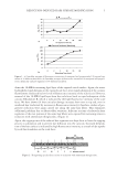
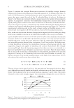
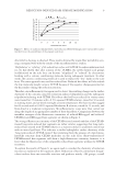
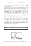
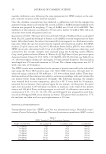
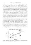
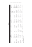
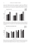
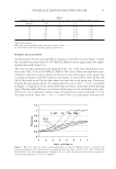
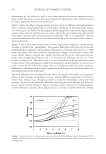
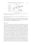
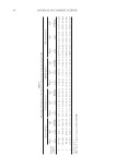
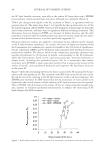
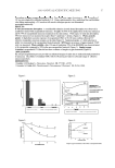
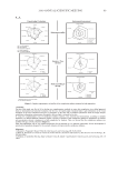
JOURNAL OF COSMETIC SCIENCE 46 the SC lipid lamellar structure, especially at the surface SC layers (fi rst strip). EPDME post-treatment clearly restored these structures, although not completely (Figure 4). TEWL also changed with depth in the SC, as shown in Table I, in agreement with our previous data (2). The values were from 7 to 8 (g/m2h) for the top layer and 14 to 19 for the fourth stripping. All the TEWL values are well within the normal range for skin with intact barrier function, and there were no signifi cant differences among treatments. This discrepancy between changes in TEWL, as a measure of barrier function, and the order parameter S indicates that the lamellar lipid structures are not the single decisive deter- minant of skin barrier function, as we have previously suggested (2). Stepwise regression analysis was applied to investigate factors infl uencing the changes in the S value between treatments. As a result, the extent of hydration measured with the Corneometer was confi rmed as a predictor variable at the 10% level of signifi cance. On the other hand, TEWL and the hydration value measured with the Skicon were not predictively valuable. The Skicon, based on the conductance principle, measures very superficial hydration (to 20 microns of depth), corresponding to the SC hydration, while the Corneometer, based on the capacitance principle, measures hydration at deeper levels, including the epidermal region (20). It is noteworthy that surface treatment with EPDME, a large-molecular polymer that is expected to remain at the surface of the SC, alters epidermal hydration and improves the lipid-layer structure in the outer SC. Figure 7 shows the relationship between the water content of the SC measured by Corne- ometer and order parameter (S). The treatment with SDS leads to the loss of water in the SC and the loss of the ordering in the SC lipid structure, both in all four strippings. The EPDME post-treatment for SDS-treated skin led the restoration of water content even higher than the control (water), and the ordering of SC lipid structures was also repaired. Hence EPDME, which remains on the surface of skin as a large-molecular-weight poly- mer, assumes to stimulate epidermal moisturization to enhance the structuring of SC lipid disrupted by SDS treatment. Figure 7. Relationship between the water content of SC measured by Corneometer and order parameter (S).

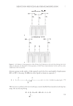
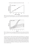
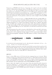
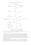
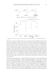
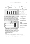
EPDME IMPROVES LAMELLAR LIPID STRUCTURE 47 To evaluate further the effect of EPDME on lipid membranes, we conducted experiments using liposomes. There are many reports of EPR studies on the fl uidity of liposome mem- branes (14,21–24), and the spectra that we obtained with 5-DSA were consistent with these reports (Figure 5). It was confi rmed that, although the lipid membrane structure was disrupted by SDS (the fl uidity is increased, as indicated by a decrease in S of ca. 0.007), pre-treatment with EPDME suppressed this effect (the reduction in S was only 0.003), as shown in Figure 6. Mizushima et al. (16) examined the moisture retention capacity of SC under various con- ditions and suggested that water in untreated SC hydrates the lipid layers and maintains the S value even under very dry external conditions. On the other hand, the amount of water in SC treated with SDS varied, depending on environmental humidity, with corre- sponding changes in the S value (16). It can be speculated that SDS disrupts lamellar structures by solubilizing the lipid components, releasing water of hydration as free wa- ter. Thus, the improvement in membrane structural order by EPDME in SDS-treated SC and liposomes in this study may be attributable to the ability of EPDME to provide bound water at the lipid layers. CONCLUSIONS EPR spectroscopy was used to examine changes in the order parameter of lipid structure in SC successively stripped from SDS-treated dry skin. Application of EPDME partially reversed the disruption of lipid structure due to SDS treatment. Similar improvement was found in SDS-treated liposomes. The low concentration of SDS used did not result in any decrease in TEWL, indicating that EPDME may preserve the ordered lamellar struc- ture of lipids by increasing the level of hydration. ACKNOWLEDGMENTS We thank Professor Noriyuki Kumazawa of Ibaraki University for providing liposomes, and Dr. Koichi Nakagawa of Fukushima Medical University and Dr. Mitsuhiro Denda of the Shiseido Research Center for helpful suggestions. REFERENCES (1) T. Ohmori, Y. Yamamura, K. Nakahara, R. Miyahara, K. Hosokawa, K. Maruyama, T. Okamoto, and H. Kakoki, Development of novel multifunctional cosmetic raw materials and their application. I. Characterization of a random copolymer of polyoxyethylene/polyoxypropylene dimethyl ether, J. Oleo Sci., 55, 365–375 (2006). (2) E. Yagi, K. Sakamoto, and K. Nakagawa, Depth dependence of stratum corneum lipid ordering: A slow-tumbling simulation for electron paramagnetic resonance, J. Invest. Dermatol., 127, 895–899 (2007). (3) E. Yagi, K. Nakagawa, and K. Sakamoto, Establishment of ex vivo stratum corneum lipid ordering analysis by electron spin resonance, J. Soc. Cosmet. Chem. Jpn., 42, 231–236 (2008). (4) J. Mizushima, Y. Kawasaki, M. Ino, K. Sakamoto, M. Kawashima, and H. I. Maibach, Effect of surfac- tant on human stratum corneum utilizing electron paramagnetic resonance spectroscopy, J. Jpn. Cosmet. Sci. Soe., 25, 130–135 (2001). (5) K. Nakagawa, J. Mizushima, Y. Takino, K. Sakamoto, and H. I. Maibach, Chain ordering of stra- tum corneum lipids investigated by EPR slow-tumbling simulation, Spectrochimica. Acta Part A, 63, 816–820 (2006).
Purchased for the exclusive use of nofirst nolast (unknown)
From: SCC Media Library & Resource Center (library.scconline.org)