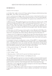
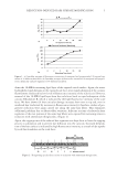
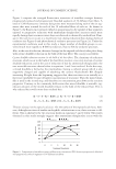
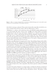
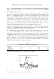
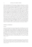
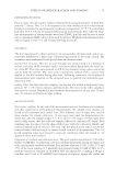
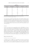
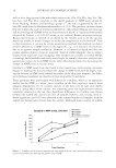
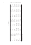
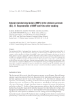
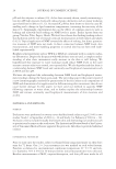
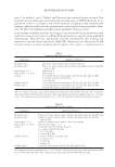
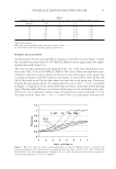
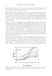
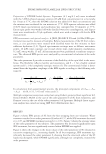
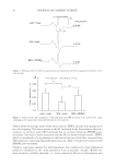
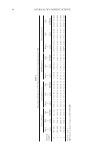
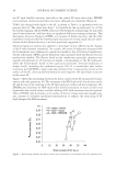
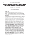
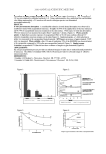
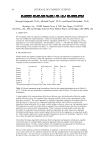
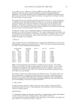
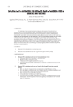
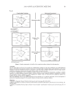
JOURNAL OF COSMETIC SCIENCE 42 Table I shows the average values of the water content, TEWL, and the order parameter S, for each stripping. The water content in the SC, measured with a Corneometer, showed a tendency to decrease upon SDS treatment but to increase following EPDME post- treatment. The water content measured with the Skicon showed similar results. TEWL, which is considered to be a parameter of the barrier function of the skin, increased after SDS treatment, indicating impaired barrier function, while it tended to be reduced with EPDME post-treatment. Stepwise regression analysis by null hypothesis was conducted to find influential predictor valiable(s) to the order parameter S as an outcome variable. Within the three predictor valiables analyzed, i.e., water content by Skicon or Corneometer and Figure 2. EPR spectrum of 5-DSA incorporated into SC lipids from the fi rst stripping of a volunteer’s mid- volar forearm. Figure 3. Values of the order parameter S obtained from the EPR spectrum of SC removed by single strippings with cyanoacrylate from the forearms of ten volunteers.

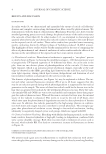
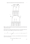
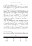
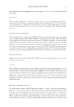
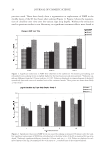
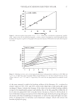
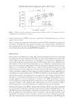
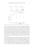
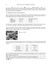
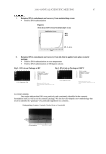
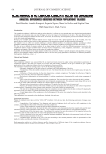
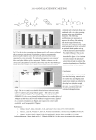
EPDME IMPROVES LAMELLAR LIPID STRUCTURE 43 barrier disruption by TEWL, only water content by Corneometer showed a level of significance of 0.099. Figure 5 shows typical spectra of liposomes treated with SDS, and Figure 6 summarizes the results. Although the order parameter S shows only a slight change, and SDS treat- ment signifi cantly increased the fl uidity of the membranes, this increase was suppressed by treatment of the liposomes with EPDME beforehand. DISCUSSION Skin cleansers are widely used to keep skin clean and sanitized, but excessive cleansing can remove natural moisturizers and SC lipids. It is also well established that surfactants, such as SDS, cause skin irritation (8–11). Imokawa et al. (8) reported a signifi cant de- crease in SC hydration in skin treated with 5% SDS, though there was no infl ammation. The amount of released SC lipid constituents, such as cholesterol, cholesterol ester, and fatty acids, increased time-dependently, leading to disruption of lamellar lipid structure. Ohmori et al. (1) applied 5% SDS solution to the mid-volar forearm for ten minutes and observed a reduction of SC hydration and barrier function. We used 1% SDS solution in order to investigate changes in lipid structure in the SC without causing signifi cant bar- rier disruption, as determined by means of TEWL and SC hydration measurements (Table I). Electron microscopy is a powerful tool to investigate structural changes in SC lipid layers directly, e.g., in skin obtained by biopsy (12), and the stripping technique has been suc- cessfully applied to electron microscopic evaluations (13). EPR measurement using ex vivo SC obtained by surface biopsy with cyanoacryrate is also suitable to examine depth- dependent changes in the ordering profi le of SC lipid layers (2). An advantage of EPR is that the operation is very simple and easy in comparison to electron microscopy. Following the application of EPR to measure the membrane fl uidity of model compounds (14), its use has recently been extended to the structural analysis of human SC (15–17). The EPR spectrum of hydrophobic spin probes incorporated into a lipid membrane re- fl ects the properties of the membrane, and it is sensitive to the rotational mobility of the spin probes, the polarity of the environment, and the orientation of the probe molecules. The order parameter (S) is determined from the hyperfi ne couplings of the EPR signals Figure 4. Values of the order parameter S obtained from the EPR spectrum of SC removed in successive strippings from the forearms of ten volunteers.
Purchased for the exclusive use of nofirst nolast (unknown)
From: SCC Media Library & Resource Center (library.scconline.org)