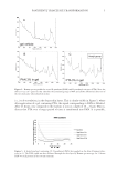
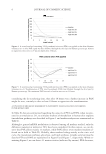
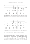
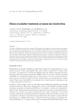
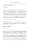
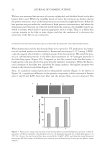
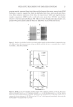
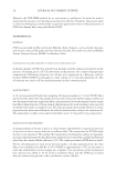
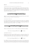
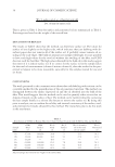
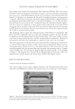
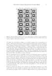
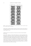
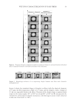
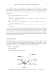
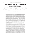
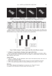
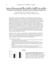
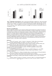
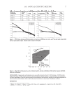
PANTHENYL TRIACETATE TRANSFORMATION 9 STIMULATION OF WOUND HEALING BY D-PANTHENYL TRIACETATE AND D-PANTHENOL The results of clinical study on wound healing in a group of human volunteers are de- scribed in Figure 8. As can be seen, transepidermal water loss (TEWL) dramatically in- creases 30 minutes after wound induction, and treatment with the different products (placebo, PTA 3%, PAN 3%) does not modify the TEWL when compared to a saline treatment. After 48 hours of treatment, all treatment groups produced a statistically signifi cant effect compared to saline (see Statistical Analysis, p. 4). However, after 72 hours of treatment with the products, only PTA was statistically signifi cant when com- pared to saline (p0.05, Student’s t-test), with a difference of -8.7%, while placebo and PAN treatments were not. Figure 7. Metabolism marker mRNAs from human skin explants treated with an emulsion containing D-panthenol (PAN) at 2%. Treatment was for six hours and 24 hours. Arrows indicate signifi cant changes. Figure 6. Metabolism marker mRNAs from human skin explants treated with an emulsion containing D-panthenyl triacetate (PTA) at 2%. Treatment was for six hours and 24 hours. Arrows indicate signifi cant changes.

JOURNAL OF COSMETIC SCIENCE 10 DISCUSSION This paper examines some activities of vitamin B5 derivatives such as D-panthenol (PAN) and D-panthenyl triacetate (PTA), with particular attention to their effect on metabolism markers and their capacity for wound-healing stimulation. In this paper, we have shown that both PAN and PTA penetrate the human skin when carried by a gel or by an emul- sion and that both elicit metabolic changes. In particular, utilizing Raman spectroscopy, we demonstrated that both PTA and PAN can travel through the stratum corneum, while PTA is slowly de-acetylated in PAN with time. Interestingly, in human volunteers this effect peaks at around 24 hours. When a human skin biopsy is observed, the metabolic changes induced by PTA start happening more rapidly (six hours). This could be due to the faster penetration of the actives in human skin explants compared to in vivo skin (14) and possibly also to the vehicle used to carry the actives (emulsion vs gel), allowing an earlier bioavailability of PTA for de-acetylation in the human skin explants. Furthermore, Raman studies show that, indeed, most PTA is converted in PAN, since almost no PTA residue is seen after 24 hours (Figure 4). If most, if not all, PTA is converted in PAN over time, it is possible that the metabolic effect observed in human skin explants (see Table II) are mostly due to PAN. Analysis of the data from the metabolic marker study indi- cates, in fact, strong analogies between PTA and PAN treatment. Both ingredients push the citric acid cycle by stimulating mitochondrial enzymes such as aconitase 2 mi- tochondrial, aconitate hydratase (ACO2), while PTA also stimulates malate dehydroge- nase 2 (MDH2). The effect on ACO2 and MDH2 could be explained, considering that panthotenic acid is a key component of coenzyme A (CoA) and that acetylCoA is upstream and feeding the citric acid cycle (see Figure 1). The same observation can be made when markers involved in the metabolism of isopren- oids or lipids, which are stimulated by PTA and PAN, are involved, with particular interest in cholesterol synthesis and modifi cation. Cholesterol synthesis is essential for homeosta- sis of the epidermis, being required for both cell division and for differentiation, as well Figure 8. Wound healing measured by transepidermal water loss (TEWL) on the skin of human volunteers (n = 37) after treatment with an emulsion containing 3% D-panthenyl acetate (PTA) or D-panthenol (PAN). T0 is 30-minute treatment after wound induction. *p0.05 vs saline, Student’s t-test.
Purchased for the exclusive use of nofirst nolast (unknown)
From: SCC Media Library & Resource Center (library.scconline.org)