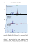
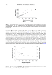
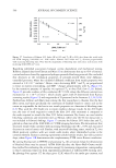
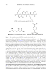
518 JOURNAL OF COSMETIC SCIENCE to the detection of saponins in other types of personal care products, which contributes to quality control throughout the efficacy personal care industry. ACKNOWLEDGMENT This study was supported by YUNNAN BAIYAO GROUP CO., LTD, Yunnan Province, China. There are no conflicts of interest for all authors. REFERENCES (1) Z. H. Y. Wu, Yunnan Flora (Science Press, Beijing, 2006), p. 509. (2) H. T. Pang, D. S. H. Luo, and J. Guo, Analysis of chemical constituents of Panax notoginseng and network pharmacological study on its anti-inflammatory mechanism, Chin. Tradit. Herb Drugs., 51, 139−148 (2020). (3) T. K. Yun, Y. S. Lee, H. Y. Kwon, and K. J. Choi, Saponin contents and anticarcinogenic effects of ginseng depending on types and ages in mice, Acta Pharmacol. Sin., 17, 293–298 (1996). (4) J. Hao, H. Hu, J. Liu, X. Wang, X. Liu, J. Wang, M. Niu, Y. Zhao, and X. Xiao, Integrated metabolomics and network pharmacology study on immunoregulation mechanisms of panax ginseng through macrophages, Evid. Based Complementary Altern. Med., 2019, 1–14 (2019). (5) X. Chen, Cardiovascular protection by ginsenosides and their nitric oxide releasing action, Clin. Exp. Pharmacol. Physiol., 23, 728–732 (2010). (6) T. Z. Tao, F. Chen, L. L. Bo, Q. Xie, W. J. Yi, Y. Zou, B. Hu, J. Li, and X. Deng, Ginsenoside Rg1 protects mouse liver against ischemia–reperfusion injury through anti-inflammatory and anti-apoptosis properties, J Surg Res., 191, 231–238 (2014). (7) M. M. Wang, M. Xue, Y. G. Xu, Y. Miao, N. Kou, L. Yang, Y. Zhang, and D. Z. Shi, Panax notoginseng saponin is superior to aspirin in inhibiting platelet adhesion to injured endothelial cells through COX pathway in vitro, Thromb. Res., 141, 146–152 (2016). (8) C. Yu, C. Diao, W. Xia, and B. Zhou, Determination of ginsenoside Rg1 in Shenfu injection by RP-HPLC, Chin. J. Pharm. Anal., 20, 263–265 (2000). (9) H. C. Wu, H. M. Liu, J. Bai, Y. Lu, and SH. Y. Du, Simultaneous determination of notoginsenoside R1, ginsenoside Rg1, ginsenoside Re and 20(S) protopanaxatriol in beagle dog plasma by ultra high performance liquid mass spectrometry after oral administration of a Panax notoginseng saponin preparation, J. Chromatogr. B Analyt. Technol. Biomed. Life Sci., 974, 42–47 (2015). (10) X. Y. Li, J. G. Sun, G. J. Wang, H. P. Hao, Y. Liang, Y. Zheng, B. Yan, and L. Sheng, Simultaneous determination of panax notoginsenoside R1, ginsenoside Rg1, Rd, Re and Rb1 in rat plasma by HPLC/ ESI/MS: platform for the pharmacokinetic evaluation of total panax notoginsenoside, a typical kind of multiple constituent traditional Chinese medicine, Biomed. Chromatogr., 21, 735–746 (2007). (11) Y. Tian, Y. Lu, J. Xie, Y. Cheng, R. Qi, Y. Wu, and S. Zhang, Rapid determination of ginsenoside Rg1, Re and Rb1 in ginseng samples by capillary electrophoresis, Anal. Methods., 1, 203 (2009). (12) Y. J. Kim, S. H. Han, J. Y. Jeon, M. H. Hwang, Y. J. Im, S. Y. Lee, S. W. Chae, and M. G. Kim, Validation of LC-MS/MS method for determination of ginsenoside Rg1 in human plasma, Anal. Sci. Tech., 26, 221–227 (2013). (13) M. S. Bispo, E. S. D. B. Morte, M. D. G. A. Korn, L. S. G. Teixeira, M. Korn, and A. C. S. Costa, Determination of Pb in river water samples by inductively coupled plasma optical emission spectrometry after ultrasound-assisted co-precipitation with manganese dioxide, Spectrochim. Acta B., 60, 653–658 (2005). (14) J. Zhou, J. Zhao, H. Yuan, Y. Meng, Y. Li, L. Wu, and X. Xue, Comparison of UPLC and HPLC for determination of trans-10-hydroxy-2-decenoic acid content in royal jelly by ultrasound-assisted extraction with internal standard, Chromatographia., 66, 185–190 (2007).

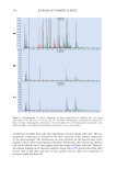
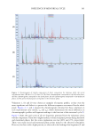
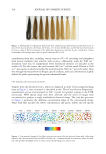
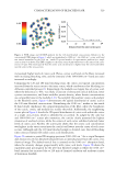
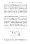
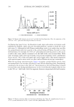
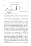
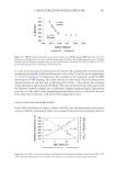
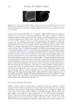
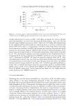
519 Address all correspondence to Timothy Gillece, tgillece@yahoo.com Characterization of Bleached Hair: Vibrational Spectroscopy, Thermal Analysis, and Determination of Equivalent Damage Factor TIMOTHY GILLECE, LARRY SENAK AND ROGER L. MCMULLEN Ashland LLC, Bridgewater, New Jersey, USA (T.G., L.S., R.L.M) Accepted for publication May 27, 2021. Synopsis In this study, we sought to determine a practical correlation between disulfide bond oxidation and the thermal response of chemically bleached hair fibers. Bleaching processes, and the alkaline environment under which they are applied, cause scission of native covalent cystine cross-links in virgin hair fibers to form cysteine-sulfenic, cysteine-sulfinic, and cysteine-sulfonic (cysteic) acids in the cuticle, cortex, and, to a lesser extent, in the medulla. To further our understanding of hair bleaching kinetics, results from Fourier transform infrared (FTIR) chemical imaging, FTIR-attenuated total reflectance (FTIR-ATR), and Raman spectroscopic measurements were correlated with results from high pressure differential scanning calorimetry (HPDSC), dry differential scanning calorimetry (DSC), dynamic vapor sorption (DVS), and modulated thermogravimetric analysis (MTGA). Spectroscopic results were used to calculate an equivalent damage factor (EDF), which was used to index bleaching damage to the cuticular and cortical compartments of the hair fiber. Spectrofluorescence and colorimetry measurements were performed on bleached whole fiber hair tresses. Fluorescence measurements provided a means to monitor changes in the tryptophan and kynurenine levels, and colorimetry measurements were conducted to quantify the overall color change (ΔE) of hair at various bleaching intervals. FTIR imaging showed that cysteic acid levels in the fibers increased with increasing bleaching time and that the spatial distribution of cysteic acid builds from the outer cortex to the inner cortex, which further validates that bleaching is a diffusion-controlled process. FTIR- ATR studies with whole fiber hair tresses and 3-µm cross-sections showed that the cuticular cysteic acid concentration changes abruptly, whereas conversion of cortical cystine to cysteic acid is diffusion limited. Raman spectroscopy perfectly complemented FTIR-ATR and FTIR imaging, in which case Raman was used to directly follow changes in cystine (509 cm−1) as a function of bleaching time, whereas FTIR spectroscopy monitored increases in cysteic acid concentration (1040 cm−1). The cortical EDF values for Raman and FTIR spectroscopic techniques correlated linearly (R2= 0.93–0.99), whereas the association between whole tress and cortical EDF results was poor (R2= 0.61–0.73). For the series of bleached fibers, changes in the denaturation temperature (TD) from HPDSC analyses obeyed Fick’s laws of diffusion (R2= 0.99), where the diffusion constant was estimated to be 1.1 × 10⁻⁸ cm2min−1. Using the peak in TD, the model-free Ozawa method was applied to approximate changes in the activation energy of intermediate filament denaturation as a function of increasing bleaching time. After 90 min of bleaching, the HPDSC activation energies plateaued at 180 ± 8 kJ/mol against increasing cysteic acid concentration. Dry DSC results showed that conversion of cystine to cysteic acid increased the cortical mobility temperature, advocating that ionic and hydrogen-bonded networks stabilized components of the dry cortex during excessive heating. The MTGA pyrolysis onset temperatures ranged from 237°C to 248°C for virgin and 240 min bleached hair tresses, J. Cosmet. Sci., 72, 519–546 (September/October 2021)
Purchased for the exclusive use of nofirst nolast (unknown)
From: SCC Media Library & Resource Center (library.scconline.org)