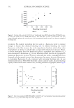
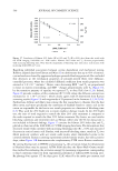
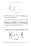
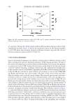
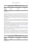
520 JOURNAL OF COSMETIC SCIENCE respectively, where the onsets positively and linearly correlated with increases in cysteic acid concentration (R2= 0.95) however, the activation energy for pyrolysis of dry fibers showed a curvilinear correlation with Raman EDF, with a peak activation energy (554 ± 9 kJ/mol) corresponding to 60–90 min bleaching times. To establish connections between water management properties and cystine oxidation, linear trends in denaturation temperature against the normalized Raman cystine band at 509 cm−1 demonstrated that decreased cross-link density is directly connected to greater steady-state moisture regains (R2= 0.94). For hair tresses, low EDF correlated with high tryptophan levels however, with increased bleaching, tryptophan and cystine levels rapidly decreased. As expected, longer bleaching times produced increased differences in color, as indexed by ΔE. INTRODUCTION Hair bleaching is one of the most common chemical treatments of hair. This cosmetic procedure is carried out to lighten hair that is naturally dark. Therefore, the objective of such treatments is to destroy and remove melanin granules from the hair fiber. This is an oxidative process that is typically achieved with hydrogen peroxide (H 2 O 2 ) and ammonia. Since melanin granules are in the cortex of hair, hair bleaching formulations must penetrate the fiber integument. Unfortunately, bleaching of hair results in collateral damage to lipids, proteins, and other structural components of the fiber. Regarding oxidative susceptibility, some of the most labile amino acids in keratin are methionine, tyrosine, threonine, and tryptophan. Even more susceptible to bleaching damage are cystine residues, which predominantly undergo oxidative fission of sulfur-sulfur (-S–S-) cross-links, resulting in the formation of salts of sulfur acids, including sulfenic acid (-SOH), sulfinic acid (-SO 2 H), and sulfonic acid (-SO 3 H). Most of the species are intermediates while sulfonic acid, which is frequently referred to as cysteic acid, is the predominate moiety remaining after normal bleaching cycles (1). The oxidation of disulfide bonds in hair has a significant influence on the various components of the fiber. In fact, the highest concentration of disulfide bonds in the hair fiber is in distinct lamellar layers of cuticle cells including the A-layer and exocuticle however, in comparison to the cortex, the cuticle does not significantly contribute to the mechanical properties of hair since it is only a fraction of the total cross- sectional area. The cortex of hair is comprised of elongated cortical cells that are separated by a cell membrane complex. The cells are filled with macrofibrils containing low-sulfur intermediate filaments (crystalline phase) embedded in an amorphous matrix of cystine- rich proteins. Therefore, major targets of bleaching damage in the cortex are the disulfide bonds of the amorphous phase proteins. Measurements including tensile strength, fatigue testing, vibrational spectroscopy, liquid retention, DSC, thermogravimetric analysis (TGA), scanning electron microscopy, and amino acid analysis, have been extensively used by researchers to survey chemical and structural damage to hair fibers as a function of alkaline bleaching treatments (2–19). In one of the earliest studies, Edman and Marti provided empirical evidence that hydrogen peroxide degrades disulfide bonds and diminishes the work required to stretch fibers (5). The investigators performed their tensile studies in distilled water to exclusively evaluate the influence of disulfide cross-links on the fiber modulus and to eliminate strength and resilience contributions from hydrogen and ionic bonding. Specifically, tensile testing provides modulus and breaking strength values for the hair fiber, where the overall hair fiber modulus involves contributions from hydrogen bonds, ionic bonds, and disulfide bonds (6). With this understanding, Robbins evaluated the wet tensile properties of bleached fibers from a single source and found that the wet tensile strength decreased by nearly 60% for

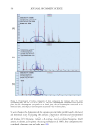
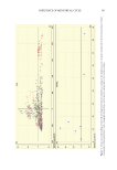
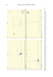
521 CHARACTERIZATION OF BLEACHED HAIR highly bleached fibers, where the compromised fibers were additionally found to have a 48% reduction in native cystine (1). More recently, thermal analysis and vibrational spectroscopy have been used to further the thermochemical understanding of oxidative bleaching damage. Thermal analysis techniques, specifically DSC, differential thermal analysis, and TGA, have been extensively employed to probe melting and pyrolysis mechanisms in keratinous materials (6–14). In dry DSC experiments, snippets of dry hair fibers are heated to 250°–300°C and the intermediate filaments are consequently denatured and pyrolyzed in a coupled thermal event. Comparatively, in HPDSC experiments, water is added to the volume of hair fibers in hermetically sealed crucibles, wherein heating the cortex in excess water plasticizes the matrix and denatures the intermediate filaments at lower temperatures (e.g., 130°–155°C) than pyrolysis events (e.g., 210°C). Because the wet-matrix viscosity and associated cystine cross-links are directly related to denaturation temperatures measured in wet DSC, HPDSC provides a means to quantify the covalent cross-link density of the matrix (6,13). Vibrational spectroscopy has also been routinely used for studying oxidative hair damage and is typically less invasive and labor intensive than tensile testing protocols. Techniques including FTIR spectroscopy, FTIR imaging, and Raman spectroscopy have been applied to evaluate increases in cysteic acid, whereas confocal Raman spectroscopic imaging has been used to chemically image complementary decreases in disulfides bonds (16–18). In the present work, results from FTIR chemical imaging, FTIR-ATR, and Raman spectroscopic measurements were compared with results from HPDSC, dry DSC, DVS, MTGA, colorimetry, and spectrofluorescence analyses to discern fundamental chemical structure/physical property relationships in chemically bleached hair fibers. Spectroscopic results were then leveraged to calculate the equivalent damage factor (EDF), which was applied to comparatively index bleaching damage in the cuticular and cortical compartments of the hair fiber. MATERIALS AND METHODS European dark brown and natural white hair tresses were bleached for 15, 30, 45, 60, 90, 120, and 240 min. Cysteic acid formation in peroxide-bleached whole fibers and cryotomed cross- sections was monitored using Raman and two FTIR spectroscopic techniques. In Raman analyses, the EDF was observed by following trends in the symmetric sulfonate (S = O) to phenylalanine ratio (1040/1003 cm−1) and the attenuation in cystine (505–510 cm−1) via the 509/1003 cm−1 band area ratio. Similar approaches were taken in FTIR-ATR spectroscopy and FTIR imaging, where the ratios of symmetric sulfonate (1040 cm−1) to the cystine monoxide stretching band at 1080 cm−1 or amide II band (1548 cm−1) were used to monitor cystine oxidation. In this work, both raw EDF and normalized EDF indices are reported. Raw EDF indices, which hereafter are referred to as EDF, were taken directly from protein- normalized cysteic acid spectral intensities, whereas normalized EDF results were evaluated to enable comparison of bleaching damage between spectroscopic techniques and band area/ intensity normalizations. Normalized EDF calculations define EDF = 1.00 for nonbleached fibers, and accordingly the EDF indices for bleached fibers were always ≥1. These data were compared to results obtained from HPDSC, where endothermic transitions such as the denaturation temperature (T D ) and denaturation enthalpy (ΔH D ) were monitored. The T D parameter correlates with changes in matrix viscosity and cross-link density, whereas ΔH D
Purchased for the exclusive use of nofirst nolast (unknown)
From: SCC Media Library & Resource Center (library.scconline.org)