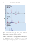
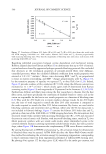
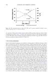
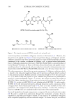
522 JOURNAL OF COSMETIC SCIENCE measures the energy required to break the interface between the matrix and intermediate filament keratin proteins (IFKPs) and the energy needed to denature IFKPs. Additionally, dry DSC, DVS, and MTGA were performed to further the understanding of bleaching kinetics and to provide correlations between keratin oxidation, cortical swellability, and cortical pyrolysis. Spectrofluorescence and colorimetry measurements were carried out on hair tresses to correlate changes in cuticular EDF with tryptophan degradation and ΔE. MATERIALS Studies were carried out on medium density European dark brown and natural white hair tresses that were purchased from International Hair Importers & Products Inc. (Glendale, NY, USA). The natural white fibers were used solely for the Raman scattering studies. The hair was supplied as large tresses constructed with rectangular pieces of wax, which secured the root ends of the hair fiber. From this large tress, ¾-in wide tresses were sampled as a function of bleaching time. Bleaching was carried out by mixing 120 g of Clairol Professional BW2 powder lightener (The Wella Corporation, Woodland Hills, CA, USA) with 147 mL of Salon Care Professional 20 Volume Clear developer (Arcadia Beauty Labs LLC, Reno, NV, USA). The resulting mixture was applied to damp hair. Bleaching was carried out for time periods of 15, 30, 45, 60, 90, 120, and 240 min. The 15, 30, 45, and 60 min bleaching experiments were accomplished using the same bleach mix and removing hair samples at selected intervals. The tresses bleached for 120, 180, and 240 min were placed in a freshly prepared bleach mix at 60, 120, and 180 min, respectively, and the 90 min sample was taken midway between the 60−120 min bleaching treatment. After each hour, the remaining tresses were thoroughly rinsed in warm tap water to remove water- soluble material prior to reimmersing the tress in fresh bleaching solution and restarting the bleaching clock. After the last bleaching step, each tress was thoroughly rinsed with 40°C tap water and then soaked in distilled water for 3 h to remove soluble leachate. The leachate removal step was repeated a total of five times for each tress. Finally, the tresses were air-dried overnight prior to subsequent analyses. HAIR CROSS-SECTION PREPARATION Quarter-inch wide excised samples of the tress were subjected to sectioning using a Leica CM3050 S (Leica Microsystems GmbH, Wetzlar, Germany) cryostat equipped with a high- profile sectioning head and Leica 818 high-profile cutting blades. Note that the cryostat blade was changed after each sample. For each sample, a 25-mm aluminum specimen disc was pre-equilibrated in dry ice (−78.5°C). A small section (1/8–1/4 in wide x 1−2 in length) of the ¾-in wide tress was removed from the center of the tress length. The tip end of the damp fiber section was then held straight and perpendicular against the platform of the disc. At the interface between the disc and tip end of the fiber bundle, a drop of distilled water was then added. The drop instantly crystallized, hence causing the tip end of the fiber bundle to adhere to the aluminum specimen disc. By pulling gently upward on the root end of the fibers and slowly adding water to the fiber bundle, a straight rod of ice-embedded hair was produced. Separately, each embedded sample was then mounted onto the specimen head (−30°C) of the cryostat, where the chamber temperature was equilibrated at −25°C.

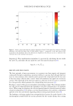
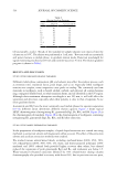
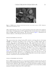
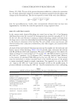
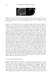
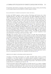
523 CHARACTERIZATION OF BLEACHED HAIR After conditioning for at least 1 h, 3- and 5-µm thick sections were collected in continuous mode using a sectioning speed of 60% maximum. Hair cross-sections were air-dried on paper overnight under reduced pressure. The cross-sections in Figure 1 demonstrate the intrinsic ellipticity of the European dark brown hair fibers. FESEM OF HAIR FIBER CROSS-SECTIONS The cross-sections were imaged using field emission scanning electron microscopy (FESEM) to determine the quality of the sections prior to FTIR imaging and to calculate the cortical cross-sectional area (n = 100). Cross-sections were fixed to aluminum PELCO® pin stubs (Ted Pella, Redding, CA, USA) using 25-mm conductive carbon tabs, and then coated with Au/Pd using our Leica EM ACE600 sputter coater. Finally, the staged fiber cross-sections were imaged with a Hitachi SU-5000 FESEM (Hitachi High Technologies, Schaumburg, IL, USA) using various magnifications. The virgin and bleached European dark brown hair cross-sections were elliptical (see Figure 1), with average major and minor diameters of 80 ± 10 µm and 57 ± 8 µm, respectively. Each fiber contained 5 ± 1 overlapping cuticle cells, and each cuticle cell was 380 ± 90 nm thick. The total pool of analyzed hair fiber cross-sections contained a mixture of medullated and non-medullated fibers. STAGING OF HAIR FIBER CROSS-SECTIONS Prior to FTIR imaging, hair fiber cross-sections must be properly staged on 25-mm calcium fluoride (CaF 2 ) windows (Spectral Systems LLC, Hopewell Junction, NY, USA). A Thermo Fisher Scientific (Waltham, MA, USA) stereo microscope and wooden toothpick were used to move cross-sections into place. In general, approximately 30 cross-sections were carefully juxtaposed without overlap on the CaF 2 crystal surface. Figure 1. FESEM image of European dark brown hair fiber cross-sections (courtesy of W.T. Thompson, Ashland Specialty Ingredients, G.P.).
Purchased for the exclusive use of nofirst nolast (unknown)
From: SCC Media Library & Resource Center (library.scconline.org)