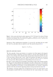
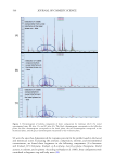
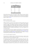
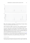
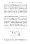
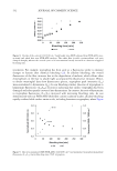
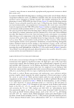
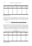
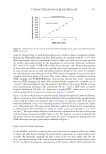
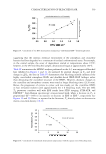
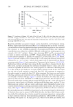
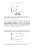
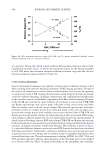
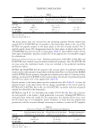
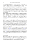
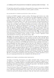
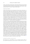
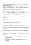
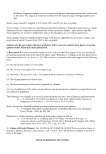
532 JOURNAL OF COSMETIC SCIENCE treatments. For example, tryptophan has been used as a fluorescence probe to monitor changes to keratin after chemical bleaching (22). In alkaline bleaching, the overall fluorescence of the fiber increases due to the degradation of melanin, which allows other chromophores in the hair to absorb light accompanied by fluorescence emission. Hence, to obtain meaningful data from fluorescence spectra, tryptophan peak intensities (I 339 ) were normalized to kynurenine (I 440 ). In our bleaching studies, the ratio of tryptophan to kynurenine fluorescence (I 339 /I 440 ) decreased, indicating that surface tryptophan had been damaged and subsequently converted into kynurenines. In contrast, the ratio of kynurenine to tryptophan fluorescence (I 440 /I 339 ) increased with increasing bleaching time. As was demonstrated with our FTIR-ATR whole fiber cystine oxidation studies, alkaline bleaching rapidly oxidizes labile surface amino acids, including keratinous tryptophan, where Figure Figure 8. Overlay of the cortical 1040/1548 cm−1 band height ratio (EDF) obtained from FTIR-ATR cross- sectional and whole hair tress FTIR-ATR analyses. The whole fiber (cuticle) cysteine-sulfonic acid ratio changed abruptly, whereas the cortical cysteic acid concentration linearly increased as a function of applied bleaching time. Figure 9. Hair tress normalized EDF (FTIR-ATR: 1040/1080 cm−1) and normalized tryptophan/kynurenine fluorescence (I339/I440) versus bleaching time. TRP: tryptophan.

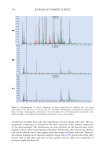
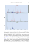
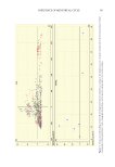
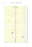
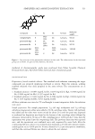
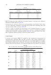
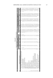
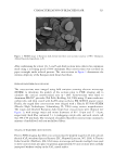
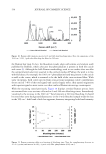
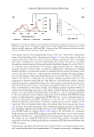
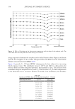
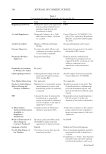
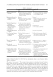
533 CHARACTERIZATION OF BLEACHED HAIR 9 reveals a steep decrease in normalized tryptophan with proportional increases in whole fiber cysteic acid. In contrast to whole fiber bleaching kinetics, oxidizing cystine in the cortical volume, which is encapsulated within the cuticle, was diffusion controlled, where the cuticular barrier initially mediated cortical absorption of alkaline peroxide. One possible explanation for the early plateauing of cortical FTIR-ATR data (first 60 min, Figure 8) is that the bleaching solution formed a viscous semisolid toward the end of each 1 h bleaching step, thereby hindering the rate of peroxide/persulfate cortical permeation. Consequently, the cuticle versus cortical EDF kinetical comparison for the FTIR-ATR studies (Figure 8 overlay), as judged by augmentation in the intensity of the normalized 1040 cm−1 cysteic acid band, is quite unambiguous. Initially, the cuticle received the brunt of the bleaching impact, whereas oxidative changes to the cortex were limited by tortuous permeation gateways introduced by cuticle and cortical diffusion networks. The disparity between the cuticle and cortical cysteic acid response is reconcilable when looking at each compartment of the hair fiber. While the morphology and molecular structure of the cuticle make it a formidable barrier for larger molecules, smaller molecules such as water, H 2 O 2 , and persulfates readily permeate the fiber structure, where diffusion control regulates further cortical permeation. Furthermore, excessive bleaching subsequently induces interfacial chemical attacks, leading to the formation of pores, cracks, and zones of erosion in the cuticle and cortex, thereby disturbing the natural diffusion processes and increasing the overall hydrophilicity of the fiber (23). As a result, trends in Figure 8 provide a “broad brush” for predicting subsequent physical properties that would be altered by cystine scission and the associated formation of cysteic acid. RAMAN SPECTROSCOPY OF CROSS-SECTIONED HAIR As the same cross-sectioning techniques for FTIR imaging and FTIR-ATR spectroscopic examinations were applied to the Raman studies, the high aspect ratio of the 3-µm thick cross-sections assured that the Raman spectra were predominantly composed of cortical and medullar shift frequencies. In the Raman experiments, instead of using European dark brown tresses, European natural white hair was employed because European dark brown hair contains melanin granules that induce excessive fluorescence, therein making it difficult to resolve Raman shifts for virgin or lightly-bleached dark brown fibers. Prior work in confocal Raman spectroscopy and multivariate curve resolution analyses provides insights toward assigning the spatial assignment of functional groups to either the cuticle or cortex (17,18). These spectral markers allow for some specificity of band assignments in the work reported by this laboratory. A table of published Raman active band assignments is presented in Table I. Works by both Kuzuhara, as well as Pudney et al., infer exclusivity of certain vibrational modes as a function of their placement in either the cuticle or cortex (17,18). Examples of such domains in the Pudney work include bands in the 650 cm−1 (-C—S-, gauche) and 880 cm−1 spectral regions of the cuticle, and conversely, the 742 cm−1 CH 2 in-phase and 1002–1003 cm−1 phenylalanine bands that prominently appear in the cortex. Of course, many vibrational bands discussed in this study are shared between physical regions, such as the 1451 cm−1 C—H bending and 1650–1675 cm−1 amide I bands. While Kuzuhara’s study agrees with Pudney on the specificity of some bands, including 1342 cm−1 (-CH 2 bend), he generally found the presence of most signature Raman bands, such as phenylalanine (1003 cm−1), in both domains of
Purchased for the exclusive use of nofirst nolast (unknown)
From: SCC Media Library & Resource Center (library.scconline.org)