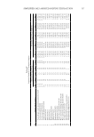
550 JOURNAL OF COSMETIC SCIENCE previously—1 M DIEA/NMP, 0.3 M HATU/DMF, 60-min reaction time, and ambient temperature. The cleavage and other processes were as described previously. In order to confirm peptide synthesis, we used an Agilent Triple Quad LC/MS 6410 mass spectrometer with an electrospray ionization interface. Peptide assay. A LC-MS/MS method for assay of KTTKS and Pal-KTTKS has been reported by Choi et al. (4). Using this approach, we developed different methods for assay of KTTKS and the conjugates by LC-MS. The concentrations of KTTKS and its derivatives were measured by a selected ion chromatogram method using Agilent Triple Quad LC/MS 6410. Capital C8-Optimal column (250× 4.6 mm, i.d. 5 µm) was used for all peptides. The mobile phase of acetonitrile and ammonium acetate solution (20 mM) containing glacial acetic acid (0.05% w/v) under isocratic elution mode was employed for all four peptides, with the proportion of 40:60 (v/v) for KTTKS, Cit-KTTKS, and Per-KTTKS and 60:40 (v/v) for Pal-KTTKS. The flow rate was 0.6 mL/min for KTTKS and Cit-KTTKS, 0.5 mL/min for Per-KTTKS, and 0.3 mL/min for Pal-KTTKS. Gas temperature and gas flow were adjusted at 350°C and 10 L/min respectively. The injection volume was 25 µL. Mass to charge (m/z) ratios of 564, 716, 712, and 802 were used for KTTKS, Cit-KTTKS, Per-KTTKS, and Pal-KTTKS respectively. Stability studies. Aqueous solutions of Cit-KTTKS and Per-KTTKS were individually prepared at a concentration of 100 µg/mL. Each solution was divided into three glass vials and placed in a 32°C water bath for 48 h. After this period of time, the concentrations of peptides were measured by LC-MS (as described previously) and they were compared to the initial concentrations. Permeation studies Membrane model permeation study. To perform this study, a lipophilic barrier was prepared by deposition of n-hexadecane into a filter using a solution of n-hexadecane in n-hexane (5% v/v) (15). PVDF (polyvinylidene fluoride) filters with pore size of 0.45 µm were mounted on the static Franz-type diffusion cells (effective surface of 3.8 cm2) and 63 µL aliquots of n-hexadecane in n-hexane solution were distributed to each filter using sampler. Diffusion cells were then placed under the fume hood, in order to evaporate the n-hexane for 1 h. After that, the aqueous solutions of different permeants (100-1000 µg/mL) were placed in the donor compartments separately. The receptor compartments were filled with deionized water. The diffusion cells were then placed for 24 h at 37°C in a water bath to have a 32°C temperature on the surface (skin temperature). Finally, the peptide concentrations in the donor and receptor phases were measured by LC-MS as described previously. We performed these experiments in triplicate. Epidermal membrane permeation study. The epidermis used in the permeation study was obtained from female donors who underwent abdominoplasty. This experiment was performed under supervision and according to ethics committee regulations of Shahid Beheshti University of Medical Sciences. After removing subcutaneous fat, skin samples were frozen at −20°C. The heat separation method was used to separate the epidermis from dermis (16). The detached epidermis was then mounted on side-by-side horizontal diffusion cells (effective surface of 1.5 cm2). The donor compartments were individually filled with aqueous solutions of peptides with concentration of 100−500 µg/mL and the receptor compartments were filled with 10%

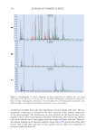
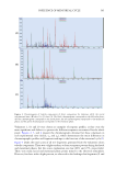
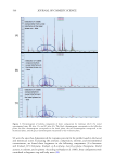
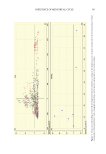
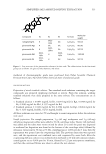
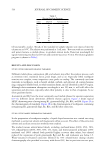
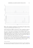
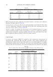
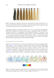
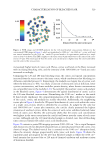
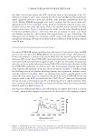
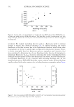
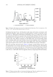
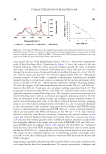
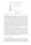
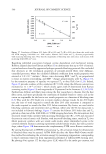
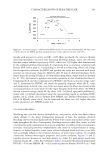
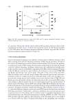
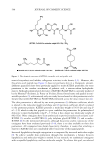
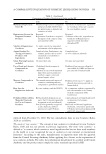
551 TERPENE CONJUGATION hydroethanolic solution. The diffusion cells were placed into a 32°C water bath and the receptor phases were stirred during the study. After 48 h, the donor and receptor phases were pulled out and analyzed. The concentrations of peptide in the donor phase as well as in the receptor phase were measured by LC-MS. We performed the experiments were performed in triplicate. RESULTS AND DISCUSSION SYNTHESIS AND CHARACTERIZATION OF PEPTIDES Cit-KTTKS, Per-KTTKS, Pal-KTTKS (Figure 2), and KTTKS (Figure 1) were synthesized by the Fmoc strategy. Using mass spectrometry, synthesis of the peptides was confirmed so that singly charged ion peak of m/z 564.2, 802.1, 715.9, and 712.1 associated with doubly charged ion peak of m/z 282.7, 401.7, 358.9, and 356.6 confirmed synthesis of KTTKS, Pal- KTTKS, Cit-KTTKS, and Per-KTTKS respectively. STABILITY STUDIES Initial concentration of peptide solutions was 100 µg/mL. This value reached 98.00 ± 3.13 µg/mL for Cit-KTTKS and 98.35 ± 1.43 µg/mL for Per-KTTKS. These results are in agreement with the stability data for KTTKS and Pal-KTTKS obtained in previous work (99.3 ± 1.7 µg/mL for KTTKS, 97.5 ± 3.6 µg/mL for Pal-KTTKS) (14). Therefore, all peptides could be considered almost stable at least in the experimental condition at 32°C for 48 h. PERMEATION STUDIES Membrane model permeation study. As described earlier, we performed the peptide permeation studies using a membrane model of PVDF-filled n-hexadecane as a lipophilic barrier. Results (Table I) showed that KTTKS and Per-KTTKS were not detected in the receptor phases, but Pal-KTTKS and Cit-KTTKS permeated the membrane in detectable amounts and showed permeation of about 4% and 1% of applied doses, respectively. Amounts of permeated peptides per area over 24 h were then normalized to concentration to obtain estimated permeability coefficient (kp). Results showed estimated kp values of 17 × 10−4 cm/h and 2.9 × 10⁻⁴ cm/h for Pal-KTTKS and Cit-KTTKS respectively. The n-hexadecane, a 16-carbon alkane, was chosen to simulate the central part of the lipid bilayer matrix of human stratum corneum (SC), an important barrier to transdermal and topical delivery of hydrophilic compounds. This barrier was used to screen peptides according to their partition coefficient (as described by Moghimi et al.) for other hydrocarbons such as isooctane (13). Results showed that Pal-KTTKS, which has the most tendency to a lipophilic environment (clog P: 3.72), had the highest estimated kp and Cit-KTTKS (clog P: -0.08) was in second place. KTTKS and Per-KTTKS that possess cLogP of less than around −1 did not permeate this lipophilic barrier. These data agree with the general consensus of skin scientists that consider LogPs of around 0–4 good for skin permeation. These data also might show that PVDF-filled n-hexadecane might be a good model for the SC permeation studies.
Purchased for the exclusive use of nofirst nolast (unknown)
From: SCC Media Library & Resource Center (library.scconline.org)