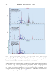
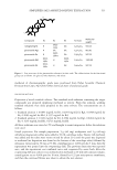
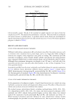
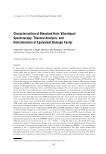
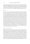
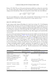
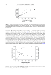
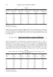
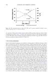
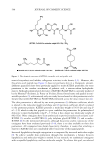
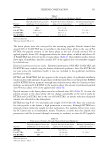
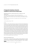
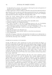
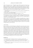
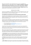
Figure 2. The chemical structures of Cit-KTTKS (citronellic acid-KTTKS), Per-KTTKS (perillic acid-KTTKS), and Pal-KTTKS (palmitic acid-KTTKS). cLogPs of conjugates were calculated by ACD-ChemSketch freeware software (). 552 JOURNAL OF COSMETIC SCIENCE

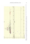
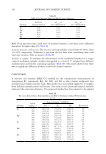
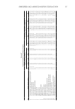
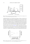
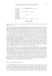
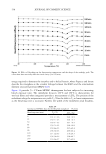
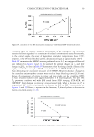
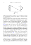
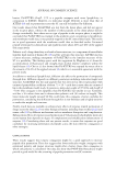
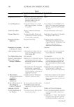
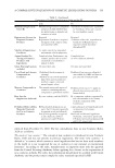
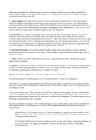
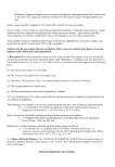
553 TERPENE CONJUGATION The donor phases were also surveyed for the remaining peptides. Results showed that around 96% of Cit-KTTKS was recoverable in the donor phase, while in the case of Pal- KTTKS, the peptide amount in the donor phase at the end of study reached 76% of applied peptide, about 24% disappearance from the donor phase, of which only about 4% of Pal-KTTKS was detected in the receptor phase (Table I). Our stability studies did not show signs of instability therefore around 20% of the applied dose was somehow trapped by n-hexadecane. Epidermal membrane permeation studies. Epidermal permeation of KTTKS, Cit-KTTKS, and Pal-KTTKS were studied using the human abdominal epidermis. Since Per-KTTKS did not pass across the membrane model, it was not included in the epidermal membrane permeation studies. KTTKS and Pal-KTTKS did not appear in the receptor phase of epidermal membrane studies in detectable amounts. In agreement with this study, Choi et al. showed that KTTKS and Pal-KTTKS did not permeate through intact hairless mouse skin (4). Contrary to these findings, we detected Cit-KTTKS in the receptor phase the amount of permeated peptide after 48 h was about 2.6% of the applied dose (Table II). Peptide amount in the donor phases was also evaluated after 48 h (Table II). As seen, the peptide amount in the donor phase did not change considerably at the end of study for KTTKS and Cit-KTTKS, but in the case of Pal-KTTKS, an intense reduction of peptide amount was observed in the donor phase. KTTKS has clog P of –3.72 and molecular weight of 563.6 Da (18). Since the action site for such peptides is the dermis, a high permeation is necessary. Although KTTKS has a relatively proper molecular weight for skin permeation, this peptide did not pass across the SC barrier due to its hydrophilic nature. Therefore, it seems that this peptide skin permeation could be increased through increasing its tendency to enter the SC lipophilic Table I Permeation of Peptides Through PVDF-Hexadecane Membrane Modela Peptide Peptide permeated across the membrane (% of initial dose) Peptide detected in the donor phases (% of initial dose) Estimated permeability coefficient (cm/h) KTTKS ND 101.4 ± 0.8 – Pal-KTTKS 3.88 ± 1.7 76.0 ± 10.0 17 × 10−4 Cit-KTTKS 0.66 ± 0.15 95.9 ± 1.7 2.9 × 10⁻⁴ Per-KTTKS ND 97.1 ± 2.9 – ND: not detected. a Data are mean ± SD, n = 3. Table II Permeation of Peptides Through Human Epidermal Membranea Peptide Peptide permeated across the membrane (% of initial dose) Peptide detected in the donor phases (% of initial dose) Estimated permeability coefficient (cm/h) KTTKS ND 102.1 ± 2.3 – Cit-KTTKS 2.64 ± 0.24 95.2 ± 0.8 7.3 × 10⁻⁴ Pal-KTTKS ND 51.7 ± 3.0 – ND: not detected. a Data are mean ± SD, n = 3.
Purchased for the exclusive use of nofirst nolast (unknown)
From: SCC Media Library & Resource Center (library.scconline.org)