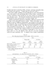
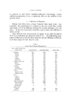
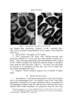
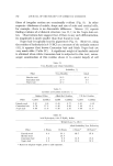
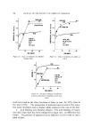
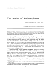
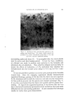
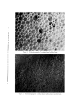
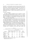
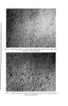
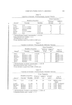
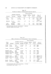
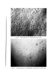
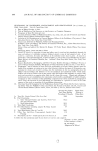
782 JOURNAL OF THE SOCIETY OF COSMETIC CHEMISTS fibers of irregular section are occasionally evident (Fig. 9). In other respects--thickness of cuticle, shape and size of scale and cortical cells, for example--there is no discernible difference. Mercer (11) reports finding evidence of a bilateral slrueture (see II, 1) in the Negro hair cor- tex. Observations here suggest that, if there is any such differentiation, its magnitude is much smaller than that found in wool. Negro hair is typically heavily pigmented (Fig. 9). However, using the residue of hydrolysis in 6/V HC1 as a measure of the melanin content (41), it appears that brown Caucasian hair and black Negro hair are very much alike (Table IV). A significant weight of insoluble material is obtained when white Caucasian hair is subjected to this test micro- scopic examination of this residue shows it to consist largely of cell Table IX Urea-Bisulfite and Alkali Solubilities Solubility, Fiber Urea-Bisulfite Alkali Lincoln wool 52.5 10.4 Caucasian hair 27.0 5.0 Negro hair 37.2 4.1 Table X Cysteine and Cystinc Contents, Original Fiber Alkali Sol. Residue UB Sol. Residue Fiber CySH CySSCy CySH CySSCy CySH CySSCy Lincoln wool 0.31 9.8 0.43 0.77 0.91 4.42 Caucasian hair 0.78 17.2 0.46 2.23 1.66 11.4 Negro hair 0.66 17.8 0.46 2.57 0.96 11.0 Table XI Acid Hydrolysis, 0.04 N H2SO4, Reflux Fiber Weight Loss (r• After) Alkali Solubility Test, Following 4 Hours 18 Hours 4 Hours* 18 Hours* Lincoln wool 0.4 21.1 82.7 97.6 Merino wool . . . 21.4 ...... Caucasian hair 1.8 6.0 18.8 74.4 Negro hair 0.2 8.0 10.5 57.4 * Based on original weight, prior to acid exposure.

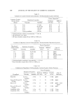
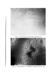
HAIR AND WOOL 783 membrane fragments. The values of the last column in the Table show that 6/V H•.SO4 leaves a similar amount of residue. One racial difference in the pigment has been reported, though on the basis of a very limited sample. Using the electron microscope, Swift (42) had measured the size of isolated melanin granules and found those from Negro hair to be larger than those from Caucasian (and also Chinese) hair. 2. Ct?emical Composition--Table V lists the results of amino acid analyses, performed by column chromatography on the three fiber samples.* Acid hydrolysates were used tryptophan, if present, was thus destroyed. Methionine was not detected in any of the samples. The results for the Lincoln wool and Caucasian hair are in general agreement with those reported by others (17). The main interest resides in the Caucasian-Negro hair comparison. Here, the only notable differences are the deficiency of serine and threonine and the excess of tyrosine, phenylalanine, and ammonia in the Negro hair. One cannot place any meaningful interpretation on these data at present. Another indication of the chemical structure is given by fractionation into "keratoses," using the method developed by Alexander (43), which is based on oxidation with peracetic acid and dissolution in aqueous ammonia. The fraction insoluble in the ammonia, which is termed •-keratose, is believed to consist of cell membranes and similar material. The fraction precipitated by the adjustment of pH down to 4, called a-keratose, is thought to originate from the crystalline portion of the protein and the residue, •,-keratose, from the amorphous protein of high sulfur content. The values for the two hair samples were determined by the pro- cedure of Corfield, Robson, and Skinner (34) the sulfur contents were determined by the oxygen combustion method of Parisot (44). The results are given in Table VI, along with those reported by Corfield et al. for Merino wool. The two hair samples fractionate almost identically. Compared with the wool, they are higher in •- and •,- and lower in a-keratose, which indicates that the hair contains a higher proportion of amorphous, high- sulfur material. The sulfur contents of all three fractions are higher in the hairs, which is indirect evidence to the effect that the fractions are themselves complex mixtures of proteins and protein degradation products. Interestingly, the proportion of the sulfur content of the * We are indebted to Dr. E. Gross of the NIAMD, NIH, Bethesda, Md., for these data.
Purchased for the exclusive use of nofirst nolast (unknown)
From: SCC Media Library & Resource Center (library.scconline.org)