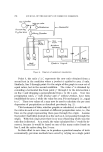
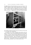
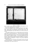
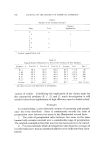
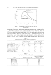
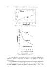
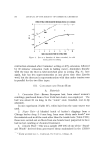
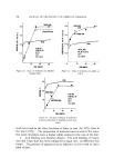
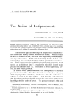
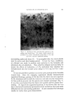
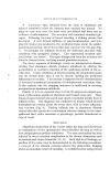
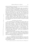
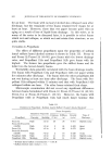
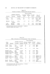
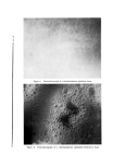
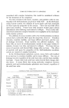
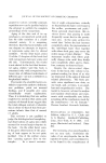
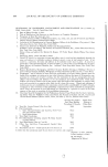
796 JOURNAL OF THE SOCIETY OF COSMETIC CHEMISTS '- •: •-*..: Figure 8. Skin biopsy from area of alu- minum salt induced anhidrosis. Ductal rupture and leakage of sweat within the epidermis produce the acute inflammatory infiltrate. Note that the superficial coils above the lesion are patent, and the duct is not dilated. Clinically these lesions resembled the prickly heat rash (H. & E. X 250) Both the dilatation of the ducts and the appearance of the PAS posi- tive nonglycogen material speak for continuing glandular function in the anhidrotic skin. Further evidence is seen by the disappearance of gly- cogen, the PAS positive diastase digested substance, from the secretory cdls. In contrast to the pre-sweat samples, those specimens taken after the forced sweating show depletion of this material. It is appropriate to comment that both the pre- and post-sweat sam- ples taken from the water treated skin sites were identical in histologic appearance to those obtained from the formalin areas, with but one im- portant exception. No discernible physical plug was present in the hydrated skin which could explain the high level blockage. This is not an unexpected result since poral closure by water is a temporary, func- tional event due to swelling of the horny layer cells at or near the eccrine ostium (11, 12).

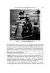
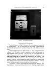
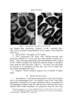
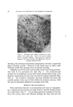
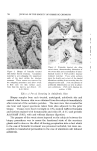
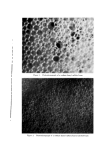
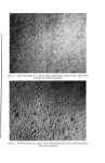
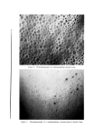
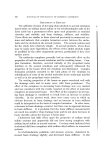
ACTION OF ANTIPERSPIRANTS 797 B. Aluminum .... Skin obtained from the areas of aluminum salt induced anhidrosis before the subjects were sweated was normal. No plugs or casts were seen, the ducts were not dilated and there was no evidence of inflammation. The secretory coil contained abundant gly- cogen. Following the hour of forced sweating, a striking picture had developed. A well formed infiltrate of polymorphonudear and lym- phocytic leukocytes localized in the periductal tissue about the epider- mal-dermal function, where the eccrine duct traverses the rete peg (Fig. 7). Occasionally the infiltrate involved the epidermis and duct wall, coincident with spongiotic changes. This represented incipient mili- arial lesions (see below). Glycogen disappeared from the secretory coil after the thermal stress, verifying normal glandular secretion. The above sequence of histologic events are interpreted as demon- strating that aluminum chloride produces anhidrosis by altering the permeability or resorptive function of the epidermal portion of the ec- crine duct. Under conditions of forced sweating the perspiration pours into the dermis faster than it can be cleared, inciting the periductal inflammatory reaction. This concept is supported by the demonstration of increased transductal permeation of iontophoresed methylene blue. It also explains why the stripping maneuver is ineffectual in restoring perspiration in aluminum anhidrosis. Finally, it is to be reported that 3 of the 20 subjects developed scat- tered erythematous papules in aluminum salt treated areas only. These lesions erupted beneath the patch and closely resembled prickly heat or miliaria mbra. This diagnosis was confirmed on biopsy, which showed intraepidermal vesicles about the sweat duct, with an acute inflamma- tory reaction (Fig. 8). Finding miliaria only in aluminum chloride treated skin also speaks for a mechanism whereby damage to the intra- epidermal duct, either anatomic or physiologic, permits transductal es- cape of sweat. DISCUSSION Superficial obstruction of the eccrine outlet has long been favored as an explanation of how spontaneous sweat disorders develop as well as how antiperspirants produce anhidrosis. The same mechanism has been indicted in sweat retention complication in the chronic dermatoses such as psoriasis and atopic dermatitis (13, 14). Such high level blockage, re- gardless of the cause, may be demonstrated by the following procedures: i, removal of the stratum corneum relieves the obstruction, bringing about an immediate return of sweating ii, occluded eccrine ostia prevent
Purchased for the exclusive use of nofirst nolast (unknown)
From: SCC Media Library & Resource Center (library.scconline.org)