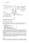
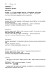
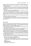
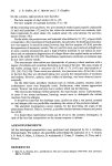
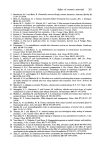
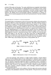
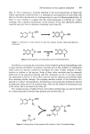
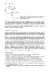
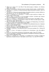
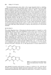
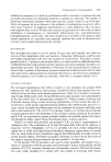
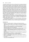
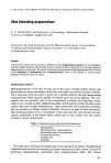
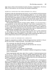
396 P. A. Riley product of the amino acid tyrosine. Two major subdivisions are recognised: pheomelanins and eumelanins. The pheomelanins are distinguished from eumelanins by their content of sulphur. They are derived from a combination of tyrosine oxidation products with cysteine which give rise to yellowish trichochromes of small molecular weight ( 1000 daltons), and reddish brown macromolecular pigments. Eumelanins, by contrast, are black or brown insoluble pigments derived from the polymerisation of tyro sine oxidation products. THE METABOLIC PATHWAY OF MELANOGENESIS The essential steps in melanogenesis consist of the enzymatic oxidation of tyrosine and its derivatives, linked to a series of spontaneous reductions. The initial oxidation consists of the addition of oxygen in the ortho position of the aromatic ring (Fig. 1). The oxidation product which is formed is 3,4-dihydroxyphenylalanine (dopa). Dopa, in common with most diphenols, readily undergoes oxidation to give rise to the corresponding quinone: the reaction consists of a dehydrogenation which is also catalysed by the enzyme tyrosin- ase. This dehydrogenation is an oxygen-requiring reaction and the oxidation of the diphenolic substrate is linked to the reduction of molecular oxygen to water. As far as the dehydrogenation reaction is concerned tyrosinase shows relatively low substrate speci- ficity with respect to the side chain. It has been shown that 5,6 dihydroxyindole is oxidised by tyrosinase and some non-physiological substrates are also oxidised, some of which (such as the hydroxylated derivatives of anisole) may have therapeutic implica- tions. Where dopa is the substrate the product is dopa quirtone (Fig. 2). co OH HO •COOH HO [0] • HO • NH 2 Figure 1. Oxidation of tyrosine to dopa. HO• CO HO OH -2H O• cOOH 0 •".• NH 2 Figure 2. Dehydrogenation of dopa to form dopa quinone. Like most quinones, dopa quinone is a highly reactive molecule, a feature which is made use of in the defensive sprays of some arthropods (1). Quinones readily condense with amino groups and will form cross-links with proteins and the reactivity of quinones may also generate undesirable deleterious effects if unrestricted access is permitted to potential substrates in the cell such as membrane lipids (see below). The spontaneous reduction steps involved in the generation of tyrosine oxidation products consist of four major reactions: reductive cyclisation, redox exchanges, reductive addition and reductive polymerisation. Indolene formation occurs by the addition of the side chain amino group to the sixth ring carbon with simultaneous reduction of dopa quinone. This gives rise to cyclo-dopa

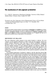
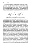
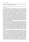
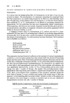
The mechanism of skb, pigment production 397 (Fig. 3). This compound is probably identical to the leucodopachrome of Raper (2). Under appropriate conditions there is a spontaneous rearrangement of cyclo-dopa with the loss of carbon dioxide and two hydrogen atoms to give 5,6 dihydroxyindole (Fig. 4). There is some evidence to suggest that this dehydrogenation is achieved by a redox exchange with a quinone intermediate. In the absence of this step alternative additive reactions may give rise to oligomers containing cyclo-dopa (3). o ••-• co OH 0 HOo•COOH H H Figure 3. Cyclization of dopa quinone to give the indolene product cyclo-dopa (leucodo- pachrome). COOH -C02 HO H -2H H Figure 4. Rearrangement of cyclo-dopa to give 5,6-dihydroxyindole. In addition to catalysing the conversion of cyclo-dopa the quinone intermediates, such as dopa quinone and indole 5,6 quinone, may take part in the oxidation of compounds which cannot be directly oxidized by tyrosinase by virtue of their structure or their location in relation to the enzyme. Some of these redox reactions may be extremely deleterious to the pigment-producing cells (for discussion see ref. 1). In man at least one mechanism is known to exist which traps the reactive quinones and prevents them from initiating cellular damage. The trapping reaction consists of the formation of a reduced C-5 adduct with glutathione. The glutamic acid and glycine residues are cleaved by peptidases giving rise to 5-S-cysteinyl dopa, whicl• can be detected in melanocytes and in the serum, and is excreted in the urine (4). The melanin group at Naples (5) have shown that cysteinyl dopa can also be formed by a direct interaction between dopa quinone and cysteine (Fig. 5). O• COOH 0 •"•"•--• N H 2 HOOC• s HOOC• SH Figure 5. Condensation of dopa quinone with cysteine to form 5-S-cysteinyl dopa.
Purchased for the exclusive use of nofirst nolast (unknown)
From: SCC Media Library & Resource Center (library.scconline.org)