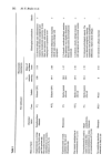
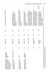
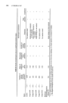
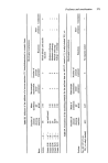
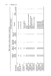
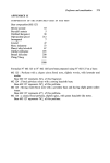
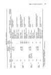
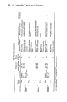
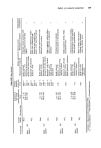
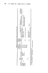
Evaluation of sensitisation potential 361 reactions. Samples are taken immediately after the readings which demonstrate these reactions. It was thought that a macroscopic examination of the lymph glands or a comparative cytological examination of the lymph glands would give a confirmation of macroscopic results. Unfortunately Freund's complete adjuvant affects all lymph ganglions modifying their form, consistency and structure and causing the appearance of pyroniphilic cells even in those ganglia furthest from the induction site. A histological examination of the skin sites was therefore found to be more fruitful. Histological exatnination After fixing fragments of skin in Bouin's solution they are embedded in paraffin, sectioned at five microns and stained with haematoxylin and eosin. When the reaction is allergic in nature the sample has the aspect of an experimental eczema. The anomalies revealed are therefore: inflammatory peri-capillary infiltration of lymphold cells with active congestion of vessels of the superficial dermis exocytosis of lymphocytes towards the epidermis and across this towards the stratum corneum intra-epidermal oedema (spongiosis) sometimes forming sub-corneal collections of liquid leading to vesicles discontinuity of the stratum granulosum surface appearance of nuclei in cells of the corneal layer (parakeratosis) between which some serous exudation is found. Later on an increase in mitotic activity of the cells of the stratum basale may occur with an increase in the number of keratinocytes of the stratum Malpighi (hyperacan- When primary irritation only is noted the alterations are different, namely: the corneal layer forms pleated folds sub-corneal eosinophilic necrosis is seen which may or may not be profound. At worst detached bubbles may develop capillary congestion of superficial dermis with polymorphic infiltration, always containing a large number of granulocytes intra-epidermal exocytosis formed only of granulocytes. However, the reaction phenomena, parakeratosis, hyperacanthosis and papillo- matosis are identical to those seen with allergic reactions. Furthermore, when the biopsy is conducted late, or when intolerance is not very marked a histological examination does not allow a definitive distinction between these two mechanisms to be made. Paradoxically it can also be difficult to distinguish between these mechanisms when intolerance is such that it results in eschar formation particularly since some substances provoke both caustic and sensitisation reactions. One should not ask too much of the classical histological examination, it has its limitations and should be regarded as one of the aids when formulating a general conclusion. Expression of results After the macroscopic cutaneous examination and histological examination have been conducted 'blind', the results are evaluated as shown below. The result is positive if one or more animals show distinct macroscopic reactions confirmed histologically as sensitisation reactions. The result is negative if no animal shows a distinct macroscopic reaction or if the histology does not confirm the macroscopic observation. The result is doubtful if a distinct macroscopic reaction is noted but the histological examination is unable to determine its origin.

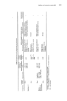
362 M. F. Brulos et al. o•o ¸ ¸
Purchased for the exclusive use of nofirst nolast (unknown)
From: SCC Media Library & Resource Center (library.scconline.org)