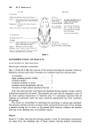
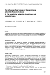
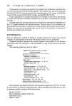
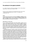
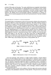
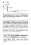
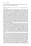
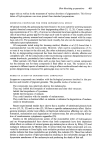
400 P. A. Riley •,,...• ' •,•..,•I 0 + Q + HzO I:'DO E + Q + H•O Figure 7. Schematic outline of catecholase activity of Neurospora tyrosinase (after Gutteridge and Robb (20). E = enzyme diphenol Q = quinone. atoms. Mason (22) furnished evidence that monophenol oxidation involves the transfer of two electrons to generate the cupric form of the enzyme. If this is confirmed it suggests that cresolase activity is consequent upon aggregation of monomeric catecholases of the type found in Neurospora and Streptomyces glaucescens (23). This leads to the interesting speculation that monophenol oxidation by tyrosinase could be controlled by factors limiting the extent of subunit interaction. MAMMALIAN TYROSINASE Studies on mammalian tyrosinase employing SDS poly-acrylamide electrophoresis by Burnet's group (24) have shown that the smallest sub-unit capable of oxidizing dopa has a molecular weight of roughly 65 000 daltons -which would correspond in size to the dimer of the Neurost•ora enzyme. In some instances another component with a molecular weight of about 120 000 daltons can also be detected which is about the size of the tetramet of the mushroom enzyme. However, a principal component in the material obtained from mice has a molecular weight of about 80 000 daltons and it is probable that this is a form of tyrosinas½ which is modified by interaction with another protein component. The struc- ture of tyrosinase is under the control of the C (or colour) locus. Modifying loci, such as pink-eyed dilution and dilute, seem to have the effect of converting more of the enzyme into the modified form. It may be that the coloration of animals carrying these genes is, therefore, modified by an interaction of tyrosinas½ with peptides which may cause a reduction in cr½solase activity, possibly as a result of a separation of the copper atoms in the dimer. Such a modification would not affect diphcnol oxidation but, by inhibiting the first step in tyrosinas½ oxidation, reduce overt pigmentation. REFERENCES 1 Riley, P. A. The mechanisms of melanogenesis. Symt•. Zool. Soc. Lond. 39 77 (1977). 2 Raper, H. S. The aerobic oxidases. Physiol. Revs. 8 245 (1928). 3 Gruhn, W. B., Pomeroy, J. S. and Maurer, L. M. An oligomeric hydroxyphenylalanine in malignant melanoma: a new type of melanogen. Biochem Biophys. res. Comm. 61 704 (1974). 4 Rorsman, H. The melanocyte illuminated. Trans. St John's Hosp. Derre. $oc. 60 135 (1974). 5 Prota, G. and Thomson, R. H. Melanin pigmentation in mammals. Endearour :t5 32 (1976). 6 Misuraca, G., Nicolaus, R. A., Prota, G. and Gliara, G. A cytochemical study of pheomelanin formation in feather papillae of New Hampshire chick embryos. ExI•erientia 25 920 (1969). 7 Geschwind, I., Huseby, R. A. and Nishioka, R. Recent Progress in Hormone Research Vol. 28, pp. 91-130, Academic Press, N.Y. (1972). 8 Commoner, B., Townsend, J. and Pake, G. E. Free radicals in biological materials. Nature 174 689 (1954).

The mechanism of skin pigment production 401 9 Mason, H. S., Ingram, H. E. and Allen, B. Free radical property of melanins. Arch. Biochern. Biophys. 86 225 (1960). 10 Pullman, A. and Pullman, B. The band structure of melanins. Biochem. biophys. Acta 54 384 (1961). 11 Van Woert, M. H. Oxidation of reduced nicotinamide adenine dinucleotide by melanin. Life Sci. 6 2605 (1967). 12 Van Woert, M. H. Reduced nicotinamide-adenine dinucleotide oxidation by melanin: inhibition by phenothiazines. Proc. Soc. Exp. Biol. Med. 129 165 (1968) 13 Gan, E. V., Haberman, H. F. and Menon, I. A. Electron transfer properties of melanin. Arch. blochem. biophys. 173 666 (1976). 14 Sarna, T., Hyde, J. S. and Swartz, H. M. Ion-exchange in melanin: An electron spin resonance study with lanthanide probes. Science. 192 1132 (1976). 15 Slater, T. F. and Riley, P. A. Photosensitisation and lysosomal damage. Nature 209 151 (1966). 16 Pathak, M. A. and Stratton, K. Free radicals in human skin before and after exposure to light. drch. Biochem. biophys. 123 468 (1968). 17 Riley, P. A. Dendritic Cells of the Epidermis. ln: Physiology and Pathophysiology of the Skin (ed. A. Jarrett) pp. 1101-1235. Academic Press, London (1974). 18 Proctor, P., McGinness, J. and Corry, P. A hypothesis on the preferential destruction of melanised tissues. J. theoret. biol. 48 19 (1974). 19 Fling, M., Horowitz, N.H. and Heinemann, S. F. The isolation and properties of crystalline tyro- sinase from Neurospora. J. Biol. Chem. 238 2045 (1963). 20 Gutteridge, S. and Robb, D. The catecholase activity of Neurospora tyrosinase. Eur. J. Biochern. 54 107 (1975). 2l Jolley, R. L., Evans, L. M., Makino, N. and Mason, H. S. Oxytyrosinase. J. Biol. Chem. 249 335 (1974). 22 Mason, H. S. The structure and functions of the phenoluse complex. Nature 177 79 (1956). 23 Lerch, K. and Ettlinger, L. Purification and properties of a tyrosinase from Streptomyces glaucescenr. Pathol. microbiol. (Basel) 38 23 (1972). 24 Holstein, T. J., Quevedo, W. C. and Burnett, J. B. Multiple forms of tyrosinase in rodents and lagomorphs with special reference to their genetic control in mice. J. exp. Zool. 177 173 (1971).
Purchased for the exclusive use of nofirst nolast (unknown)
From: SCC Media Library & Resource Center (library.scconline.org)