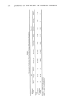
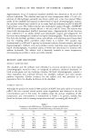
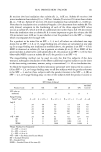
PSEUDOMONAS ON SKIN 227 swabbed, as were the finger web (between the first and second digits) and toe web (between the first and second toes). The samples obtained were serially diluted in 0.037 M PBS and pH 7.9, containing 0.05% Triton X-100, and cultured on Pseudomonas isolation agar (Difco). Plates were incubated at 37 ø C for 48 hours, and the colony-forming units were counted. P. aeruginosa was identified by appropriate biochemical methods (3). RESULTS AND DISCUSSION Four of the 166 individuals (2.5%) initially screened (subjects//5, 9, 10, and 11) carried P. aeruginosa (Table II, trial 1). One of the four subjects (//11) was negative for P. aeruginosa on three subsequent rescreenings (Table II, trials 2, 3, and 4). Subject//9 was positive for toe web carriage on initial screening, but became positive for the axilla (1 of 3 times) upon screening. P. aeruginosa had not disappeared from the toe site of this individual when the axilla was positive for this organism. When the remaining 162 subjects were resampled, seven additional P. aeruginosa carriers were detected. Of the combined total of 11 individuals who carried P. aeruginosa on the skin, seven (63%) were positive just once in the four times sampled. Of the remaining four individuals, three carried the organism twice and one carried it three times in four samplings. Those subjects from whom P. aeruginosa had disappeared repeatedly did not, however, carry the organism in the same site at each sampling. No subjects consistently carried P. aeruginosa. In no instance was P. aeruginosa recovered from the skin of nurses working either in the burn unit or in pediatrics. The ratio of P. aeruginosa skin carriage in nurses from females to males (7 females, 4 males, 64%, 36%) was approximately the same as the female to male ratio in the test population (65% females, 35% males). The mean age of individuals who harbored P. aeruginosa on their skin was 45 years (range: 26-70 years). Table II Carriage of Pseudomonas Aeruginosa at Various Skin Sites in 166 Subjects Over a 6-month Period Trial Subject a 1 2 3 4 1 -- -- toe b (40) -- 2 -- groin (350) -- -- 3 -- axilla (20) -- toe, groin (300) 4 -- axilla (10) -- -- 5 groin • (+) toe (300) toe (50) -- 6 -- -- -- axilla (200) 7 -- -- axilla (300) -- 8 -- -- -- toe (102) 9 toe (40) -- axilla (300) -- 10 groin (102) -- axilla, groin, -- (107, 800) toe (102) 11 groin, toe (+) -- -- -- aOnly subjects with P. aeruginosa at any given site are shown here. bcounts/site Ccounts not determined

228 JOURNAL OF THE SOCIETY OF COSMETIC CHEMISTS Twenty positive samples were obtained from the 11 subjects who carried P. aeruginosa. Finger web samples were consistently negative for P. aeruginosa, but the organism was detected in six instances of sampling from the axilla and six from the groin (6 to 20 samples 30%). Eight of the 20 positive samplings were from the toe web (40%). In only three instances did the subjects carry P. aeruginosa at more than one site on any given day (subject #3, trial 5 #10, trial 4 #11, trial 1). P. aeruginosa could not be recovered from the eyelids of subjects wearing contact lenses (either hard or soft). Our interest in Pseudomonas aeruginosa as a contaminant of cosmetic products is due to its pathogenicity and high environmental resiliency. This investigation was the first in which a large population of human subjects (hospital and non-hospital personnel) were examined specifically for the presence of cutaneous P. aeruginosa. Most of the earlier studies were concerned with the spread of Pseudomonas in hospital burn units (4,5). We examined intertrigenous areas because gram negative bacteria colonize in mainly warm, moist areas and are extremely sensitive to desiccation (6). Our study demonstrates that P. aeruginosa is not part of the usual microbial flora of the skin, particularly the axilla, groin, finger web and toe web. However, the bacteria may be a transient resident. Although P. aeruginosa is ubiquitous in nature, when it is experimentally applied to normal skin, its number is significantly reduced within a few hours (7). This reduction can be attributed to the inherent ability of skin to destroy certain exogenous microbes. Pseudomonal infections, related to contaminated masca- ra, probably originate from external sources rather than from skin sites. If human skin is involved, it would serve as a vehicle rather than a reservoir of this organism. This investigation was supported by a research contract from the Food and Drug Administration. REFERENCES (1) L. A. Wilson, and D. G. Ahearn, Pseudomonas induced corneal ulcers associated with contaminated eye mascaras, Am.J. Ophthalmol., 84, 112-119 (1977). (2) L. A. Wilson, A.J. Lulian, and D. G. Abeam, The survival and growth of microorganisms in mascaras during use, Am.J. Ophthalmol. 79, 596-599 (1975). (3) R. Aly and H. I. Maibach, Clinical Skin Microbiology, (C.C. Thomas, Springfield, Illinois, 1978), pp 2o-29. (4) E.J.L. Lowbury and J. Fox, The epidemiology of infection with Pseudomonas pyocyanea in a burn unit, J. Hyg. 52,403 (1954). (5) T. Rosenbury, Microorganisms Indigenous to Man, (McGraw-Hill Book Co., New York, 1962) p 126. (6) R. Aly and H. I. Maibach, Aerobic microbial flora of intertrigenous skin, Appl. Environ. Microbiol, 33, 97-100 (1977). (7) R. Aly and H. I. Maibach, In vivo methods for testing topical antimicrobial agents,J. $oc. Cosmet Chem., 32, 317-323 (1981).
Purchased for the exclusive use of nofirst nolast (unknown)
From: SCC Media Library & Resource Center (library.scconline.org)