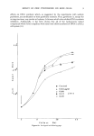
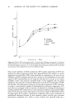
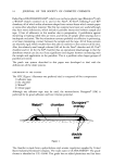
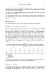
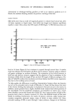
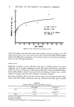
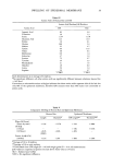
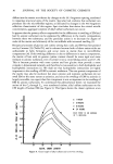
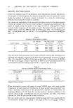
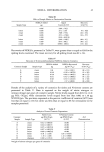
38 JOURNAL OF THE SOCIETY OF COSMETIC CHEMISTS dextrose agar acidified to pH 3.5. Most common fungal contaminants such as Penicillium and Aspergillus can be identified by colonial and cellular morphology. The more uncommon fungi require an experienced mycologist for identification and determination of any pathogenic or toxigenic potential. In addition to the media above, dilutions of talcs and powders are also plated on both anaerobe agar and blood agar. The anaerobe agar is incubated in the absence of oxygen to detect and enumerate obligate anaerobes such as Clostridium tetani, and the blood agar plates are incubated in an atmosphere containing 5% to 10% CO2 in air to detect microaerophilic organisms. ENRICHMENT AND IDENTIFICATION Final identification of contaminating microorganisms is dependent upon the results of the enrichment stage. The enrichment step is perhaps a misnomer from a strictly microbiological point of view, since the medium and conditions for incubation are not designed to enrich for specific groups of microorganisms. For example, both gram negative and gram positive organisms can be recovered. The dilutions of product (10 -• tO 10 -6) previously prepared in modified letheen broth for the plate counts are incubated at 30øC for no less than 7 days. Each dilution exhibiting growth is then streaked onto both modified letheen agar and MacConkey agar. After incubation, the plates of modified letheen agar are examined for growth, and cells for morphologically distinct colonies are gram stained. Gram positive bacilli are examined for motility and formation, morphology, and location of spores. Further characterization is usually unnecessary. Gram positive cocci are examined in somewhat greater detail. Production of catalase is determined using a solution of 3% H202. Depending on the results of the catalase test several types of media are inoculated to assist in identifying these organisms. If catalase is not produced, the following media are inoculated: a bile esculin agar slant, a tryptic soy agar slant containing 6.5% NaC1, and a blood agar plate. Blackening of the bile esculin medium along with growth in the presence of 6.5% NaCI characterizes the organism as a $treptococcus group D, enterococcus. However, if the bile esculin medium is black and there is no growth in the presence of 6.5% NaC1, the organism is a Streptococcus group D, non-enterococcus. Organisms incapable of blackening the bile esculin medium are characterized by their reaction on blood agar as either alpha, beta, or gamma hemolytic streptococci. If catalase is produced, the following media are inoculated: a mannitol salt agar plate, two tubes of oxidative-fermentative (OF) dextrose medium (one overlaid with sterile mineral oil), and an enriched agar slant (e.g., heart infusion agar) for use in the coagulase test. Ability to produce coagulase and ferment mannitol identifies the organism as Staphylococcus aureus. Oxidation and fermentation of OF dextrose without mannitol fermentation or coagulase activity characterizes the organism as Staphylococcus epidermidis. Micrococcus species are distinguished by their ability to oxidize but not ferment OF dextrose. For detection of gram negative organisms, MacConkey agar plates are streaked from the enrichment cultures each distinct morphological colony type is subcultured on triple sugar iron agar, heart infusion agar, and heart infusion broth. Because the original enrichment cultures were prepared as a series of product dilutions, it is possible to get some idea about the numbers of gram negative organisms present in the sample by

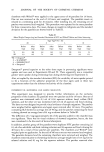
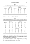
FDA METHODOLOGY FOR MICROBIOLOGICAL ANALYSIS 39 presuming that at least one organism existed in the highest dilution exhibiting growth on a streaked MacConkey agar plate. Thus, if growth occurred at the 10 -4 dilution but not at the 10 -5, one can deduce that at least 10,000 gram negative organisms per portion of sample tested (1 g or 10 g) were originally present (5). This technique, sometimes called "indicated numbers," is similar to a most probable number (MPN) method and as such cannot be compared in accuracy with the plate count method. In practice, we have frequently determined that the plate counts almost always yield a lower number of organisms. The current legal interpretation of the Food, Drug and Cosmetic Act as it applies to microbial contamination of cosmetics and topical drugs impels FDA to identify to species all gram negative bacilli for health hazard evaluation. Recently, excellent identification of both enterics as well as many non-fermenters have been attained by employing the API 20 E system as a rapid screening tool. Final confirmatory identification for the non-fermenters is still based upon the media and techniques outlined in the King scheme (6). Since for the most part, cosmetic and topical drug preparations are applied to non-sterile body areas, there is no legal requirement to manufacture a sterile product. Nevertheless, these products should be free of such pathogens as Escherichia coli, Pseudomonas aeruginosa, Staphylococcus aureus, and Salmonella and contain an effective preservative system that will ideally produce and maintain an adequately preserved product. It must be stressed, that the use of a preservative system should not in any way substitute or compensate for an unsanitary manufactured product. Presently, FDA has not set any legal microbiological limits for cosmetics and topical drug preparations and as such, each case is evaluated on an individual basis as to its public health risk (7). ACKNOWLEDGMENTS I thank James D. Macmillan, Science Advisor, Food and Drug Administration, New York Regional Laboratory, and Department of Biochemistry and Microbiology, Rutgers University, New Brunswick, NJ, and Joseph M. Madden, Food and Drug Administration, Division of Microbiology, Washington, D.C. REFERENCES (1) Bacteriological Analytical Manual, Division of Microbiology, Food and Drug Administration, Washington, D.C., 5th edition, 1978. (2) B. P. Dey and F. B. Engley, Jr., Environmental Sampling Devices for Neutralization of Disinfectants (Marion Scientific Corporation, Kansas City, Missouri, 1978), pp 1-21. (3) J. R. Evans, M. M. Gilden, and C. W. Bruch, Methods for isolating and identifying objectionable gram-negative bacteria and endotoxins from topical products, J. Soc. Cosmet. Chem., 23, 549-564 (August 1972). (4) R. S. Flowers and Z. J. Ordal, Current methods to detect stressed staphylococci, J. of Food Protection, 42, 362-367 (April 1979). (5) L. A. Otto, "Microbiology Methods for Topical Products," FDA By-Lines, 3, 209-214 (January 1973). (6) E. O. King, The Identification of Unusual Pathogenic Gram Negative Bacteria (Center for Disease Control, Atlanta, Georgia, Revised 1972). (7) J. M. Madden and G. J. Jackson, "Cosmetic preservation and microbes: viewpoint of the Food and Drug Administration," Cosmetics and Toiletries, 96, 75-77 (October 1981).
Purchased for the exclusive use of nofirst nolast (unknown)
From: SCC Media Library & Resource Center (library.scconline.org)