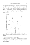
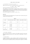
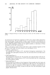
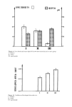
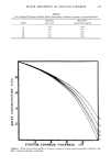
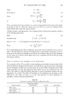
j. Soc. Cosmet. Chem., 34, 177-190 (July 1983) Skin relief and aging P. CORCUFF, J. de RIGAL, and J. L. LEVEQUE, Laboratoire de Recherche de L'Oreal, 1, avenue de Saint-Germain, 93601 Aulnay Sous Bols, France S. MAKKI and P. AGACHE, Clinique Dermatologique Universitaire, Hopital Saint-Jacques, 25030 mesancon, France. Received October 11, 1982. Synopsis An analysis using a Quantimet 720 © of skin surface negative replicas made of silicone rubber (SILFLO) © shows several modifications with age (from 2 to 98 years old) of forearm skin surface patterns of males and females. These changes of skin relief involve the principal and secondary directions of furrows, their densities and average depths, and the coefficient of developed skin surface (a measure of true versus apparent surface area). In children and adults, two main pattern directions of furrows are found. The first direction persists all through the life span, whereas the second one progressively disappears after the age of sixty and is very rarely detected beyond the age of seventy. With aging, the principal pattern direction gets closer to the forearm axis at an angle of 65 ø in children, 50 ø in adults, and 30 ø in the aged group. The skin furrow density is clearly decreased after age sixty-five, and their depths continuously increase after age fifty (children: 33 •m _+ 1 adults: 41 •m _+ 2 aged people: 60 •m _+ 3). No difference in results was found in relation to sex. INTRODUCTION The skin surface is not flat. It is characterized by a special relief which probably reproduces the three dimensional organization of the deeper layers. This skin relief structure expresses the physical state of the integument, its mechanical properties, and its possible alteration by physical factors or aging. However, a simple examination with the naked eye, or with high microscopic magnification, or with hand palpation can give qualitative information. The methods of quantitative measurements of skin microtopography have been developed in recent years (1-3). These techniques are mostly based on making negative replicas from the skin surface. The negative replicas are then used for producing positive casts which are submitted to profile analysis. The quantitative parameters used to study the skin surface 177

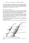
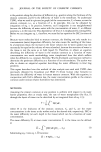
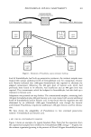
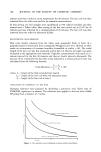
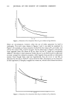
178 JOURNAL OF THE SOCIETY OF COSMETIC CHEMISTS are the same ones applied in surface engineering for measuring roughness, amplitudes, and wavelengths. Making positive replicas and numerous scans of surface profiles are time consuming. Recently, a new method using an image processing system was reported (4). We used this technique to study aging mechanism with cutaneous microtopography on 116 people from age 2 to 98 years. MATERIAL AND METHODS We chose the method described by Makki et al. (1) to obtain negative Silflo © replicas on a circular patch test of 15 mm internal diameter. A small tongue is used as a mark for alignment with respect to the body axis. Each sample (replica plus adhesive) is glued to a black metallic holder which ensures that the field to be analysed is plane. The holder is then inserted in a rotatory system graduated in degrees and centered under the plumbicon head of a Quantimet © 720 (Cambridge Instrument Co.). With a 55 mm lens a magnification of fifteen times is obtained on the TV display. Incident lighting of the replica with two floodlights generates shadows behind the furrows. These shadows are detected by their grey level. The incident lighting is opposite to the video scan direction, so intercept lines, corresponding to the horizontal projection of all detected areas, are superimposed on wrinkles and reproduce them. Field boundaries are fixed by a centered circular frame of 300 pixels in diameter. With Axsl • LIGHTING _AXS 2 Figure 1. Schematic representation of the negative skin relief and basic principle of the Image Analysis Method.
Purchased for the exclusive use of nofirst nolast (unknown)
From: SCC Media Library & Resource Center (library.scconline.org)