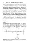
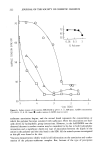
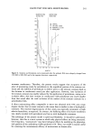
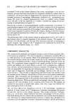
PENETRATION OF WATER AND SLS 231 •E ,- 200 o E • 1 O0 E (.1 6 12 18 24 30 36 42 48 Time (hours) Figure 2. Effect of 0.1% (A) 1.0% ([•) and 10% (O) SLS on the penetration of water. Each point represents a mean value -- SEM of five determinations (three donors). skin, possibly by extraction of lipids (10), whereby its diffusivity is changed. This is further supported by the continuous increase in penetration rate during the exposure, i.e., the absence of steady state. The similarity in p-Kp values of 10 and 1.0% SLS was unexpected. The critical micelie concentration, CMC, is 0.24% for SLS. Above the CMC the added surfactant is sup- posed to exist in the solution as micelies. These micelies do not penetrate the skin, due to the combination of size and negative charge. Hence, under infinite conditions, as in the present study, the amount penetrating from 1.0% solution should equal that pene- Table II The Apparent Penetration Rate (mg/cm-2/hr - 2) of Tritiated Water at Different Times During Exposure to Aqueous Solutions of 0.1, 1.0, and 10% (w/w) SLS Conc. 9-12 21-24 (%) (hr) (hr) 0.1 3.0 -+ 1.1 3.3 --- 1.1 1.0 10.2 _+ 3.2 7.2 _+ 1.1 10.0 10.6 ___ 3.4 7.8 _+ 1.1 Results are mean + SEM (n = 5).

232 JOURNAL OF THE SOCIETY OF COSMETIC CHEMISTS trating from 10% solution. Thus, the obtained result differs from that expected from theory. However, others have also found similar phenomenon in both dialysis (18) and ultrafiltration (19) experiments, as well as with biological materials (20,21). Thus, the monomer activity may not be constant above the CMC but may increase when the concentration of micelies increases. Above the CMC, the SLS activity seemed to be proportional to the increase in concentration (the same p-Kp for 1.0 and 10% SLS), whereas an increase from 0.1 to 1.0% SLS increased the activity much more. Thus, a practical outcome may be that skin damage from surfactants is not linearily related to the concentration of surfactant in the washing solution. The increase in permeability of skin to water appears to parallel that of SLS. The pene- tration of water was approximately the same during the exposure to 1.0 and 10% SLS, whereas a lower rate was obtained during the exposure to 0.1%. A more detailed de- scription of the correlation was difficult, because of the rapid decrease in radiochemical concentration of water in the donor solution during the exposure. This clearly shows that formulations applied to the skin can easily change composition. The penetration rate of water was in the same range as reported by other workers measuring the transepidermal water loss (TEWL) in irritation studies in viva (4, 5). The TEWL has been proposed to be a signal for recovery of the barrier function via regula- tion of epidermal biosynthesis (12,22-24). The irritation studies in viva usually involve exposure of the skin to a filter paper saturated with the SLS solution, whereafter the TEWL is measured. In these studies, values between 1.5 and 8.4 mg/cm2/hr x after exposure to 2.0% SLS were reported (4,5). In the present study the penetration rates of water were about 3 and 10 mg/cm2/hr • when 1.0 and 10% SLS were present, respec- tively. Differences in results might be due to at least two things. First, it is reasonable to expect that the degree of hydration of the skin in the present study was well above the degree of hydration in viva. In viva the TEWL is measured after equilibration of the skin to the atmosphere, whereas in the present study the diffusion of water was mea- sured during exposure of the skin to water. As the degree of hydration increases, the penetration rate also increases, since the diffusing water molecules encounter lower restraining forces (25). Second, there is much variability in permeability among indi- viduals and skin sites (3,5,14-16,26-28). In conclusion, above the CMC the influence of SLS on the barrier properties seems to be the same for 1.0 and 10% SLS, and the amount of SLS penetrating the skin was propor- tional to the applied concentration. This indicates that the monomer activity is not constant above the CMC. Below the CMC, SLS influenced the skin less, and, accord- ingly, penetrated the skin at a lower rate. ACKNOWLEDGMENTS I would like to thank Dr. Magnus Lindberg, Karolinska Institute, for valuable criticism of the manuscript, and Ms Mona Fabricius-Hansen and the other members of the staff at the Department of Plastic Surgery, Sabbatsbergs Hospital, Stockholm, for kindly supplying the skin samples. REFERENCES (1) R. Scheuplein and L. Ross, Effects of surfactants and solvents on the permeability of epidermis, J. Soc. Cosmet. Chem., 21, 853-873, 1970.
Purchased for the exclusive use of nofirst nolast (unknown)
From: SCC Media Library & Resource Center (library.scconline.org)