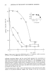
SKIN AND IMMUNITY 251 each individual will influence the response of the person to immunological stimuli and account for reduced responsiveness in aged persons because the thymus regresses with age, resulting in lowered levels of thymosin (3). The initial or primary stimulation of an immunological response is induced by antigen taken up by macrophages (phagocytes), in which much of it is degraded and excreted. A small amount persists in a modified form and is passed by direct contact between the macrophage and the T- or B-cells, which then become endowed with immunological specificity for that antigen. Langerhans cells (LC) in the skin and lymphoid tissue ap- pear to have this property of macrophages. Most antigens inducing formation of antibody are thymus-dependent and therefore T- cell-dependent. On primary stimulation there is an interaction or cooperation between T-helper cells and B-cells. The stimulated B-cells divide, and some eventually trans- form into plasma cells synthesizing and releasing antibody (4). B-cells synthesize im- munoglobulin, which remains bound on their membrane. Plasma cells derived from the B-cells form the large amounts of antibody that are released into the plasma. The immunoglobulins formed as a result of T-cell cooperation are known as IgG, IgE, and probably most of the IgA. IgG is the most important of the Ig antibodies in limiting the harmful effects of bacteria, virus, and parasites. T-cells circulate in the blood and act as "responder cells." They "recognize" foreign antigens and are stimulated by them to send signals to attract macrophages to the site (chemotaxis) for phagocytosis (engulfing of the foreign particles for example, microor- ganisms, by phagocytes). Thus, T-cells and antigens stimulate both antibody produc- tion and cellular defensive mechanisms. They do not produce antibodies per se, but they do bear specific T-cell receptors on their surface that selectively bind antigens. Like B-cells, T-cells react to antigen stimulation by secreting molecules that mediate their immune function. On the basis of the molecules they secrete, T-cells have been subcategorized as helper T-cells and cytotoxic T-cells. Today it is clear that helper T-cells fulfill their role by secreting interleukins. Cytotoxic T-cells, in contrast, make direct contact with infected cells and, by secreting toxic molecules, kill the cells and the microbes they contain (4). In addition to the helper and cytotoxic cells, the occurrence and intensity of an immu- nological response is regulated by subsets of T-cells termed suppressor cells. Suppressor cells reduce or inhibit the activity of T-cells in delayed hypersensitivity, and of B-cells in the formation of antibody. They participate in maintaining tolerance to tissue and other antigens. Failure of suppressor cell activity can result in disorders associated with T-cell abnormalities. Thus, the clinical manifestations of an immunological response are due to the activities of the effector T- and B-lymphocytes and of the suppressor cells. Allergic contact dermatitis or delayed type contact hypersensitivity (CH) to conjugates of simple chemical haptens is an example of the exquisite specificity of T-lymphocytes for small defined antigens. In general, the ability of a chemical to elicit contact sensi- tivity has been shown to directly correlate with its ability to combine with proteins. These observations led to the concept that the antigen recognized by T-cells in contact sensitivity was hapten-conjugated epidermal proteins. Several studies have shown that T-cells from contact-sensitized human or guinea pig donors could be activated in vitro by haptens conjugated to epidermal extracts, erythrocytes, or leucocyte antigens, all of which may be normal carriers for in vitro hapten sensitization. In delayed CH, antigen-

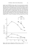
252 JOURNAL OF THE SOCIETY OF COSMETIC CHEMISTS stimulated T-cells in skin release substances that attract macrophages to the site (che- motaxis), prevent them from leaving the site (migration inhibition), enhance their ac- cumulation, and activate them for phagocytosis for increased bactericidal activity and increased secretion of macrophage inflammatory mediators (e.g., prostaglandin pro- teases). The result in a delayed hypersensitivity lesion is a compact focus of highly active cells able to kill the organisms or to render it harmless by sealing off and pre- venting its multiplication and spread (1). Psoriasis has a number of immunopathological cascades contributing to the disease. A major parameter appears to be a gene defect that leads to malfunction of the suppressor T-cells, which prevents recognition of the antigenic epidermal material. The absence of T-cell clones suppresses recognition of basal cell antigens, leading to disturbed matura- tion and impaired keratinization of basal cells, resulting in a self-perpetuating inflam- matory process (1). The predominant defect in the immune system on aging appears to be in the T-cell. In aged persons there is a general depression of the immune response. The number and function of T-cells decrease, and they lose their functional capacities in responding to specific antigens. Age changes in the T-lymphocytes are responsible for much of the pathology that accompanies aging, e.g., cancer. COMPLEMENT, PHAGOCYTES The interaction of antibodies and antigens activates a series of serum proteins collec- tively known as complement. The role of antibodies in complement-mediated effects is to confer specificity on the response. Many of the physiological activities mediated by the humoral immune system are actually carried out by the complement system. The production of antibodies provides a means of identifying foreign molecules within the body. The complement system responds to the signals provided by this recognition system and mediates one or more effector activities, such as phagocytosis, inflamma- tion, cell lysis, and the solubilization of immune complexes. The phagocytic cells of the body are responsible for ingesting and destroying particulate matter. There are two major types of phagocytes: neutrophils and macrophages. Neither of the cells are im- mune cells per se. They are able to engulf foreign particles without any assistance from the immune system. However, the specificity of their action can be greatly enhanced by antibodies. Both neutrophils and macrophages have receptors for a portion of IgG molecules. The phagocytes bind to the antibody-coated targets through these receptors, enhancing phagocytosis. While neutrophils and macrophages share many features, neu- trophils are the most abundant of the circulating white blood cells. Macrophages are localized in such tissues as lymph nodes, spleen, skin, liver, lungs, and wherever for- eign matter gains access to the body. The cell most affected by lymphocyte activity is the macrophage. Lymphocyte products, or direct contact with T-lymphocytes, stimulates macrophages that greatly increase their activity in their allergic reactions. Thus antigen-stimulated T-lympho- cytes, e.g., on the skin, release substances that attract macrophages to the site, prevent them from leaving the site, enhance their accumulation, and activate them for phagocy- tosis. The result in a delayed hypersensitivity lesion is a compact focus of highly active cells able to kill the organism, or render it harmless by sealing it off and preventing its
Purchased for the exclusive use of nofirst nolast (unknown)
From: SCC Media Library & Resource Center (library.scconline.org)