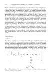
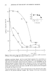
SKIN AND IMMUNITY 253 multiplication and spread (1). In addition to serving as part of the effector arm of humoral immunity, macrophages play an important part in cell-mediated immune re- sponses. The helper T-cell must receive two types of signal in order to respond to a soluble antigen. The T-cell must recognize the antigen in association with a specific macro- phage. In addition, a second signal is non-specific, provided by the cytokine or macro- phage interleukin-1 (IL-1). Thus IL-1 is a lymphocyte-activating factor produced by antigen-presenting cells (APC) in response to antigenic stimuli enhancing T-cell activa- tion (5). LANGERHANS CELLS (LC) The most compelling evidence to support a primary role for skin in immunity concerns observations made about dendritic (branch-like) bone-marrow-derived leucocytes that reside in the epidermis. These are the epidermal Langerhans cells (LC), which are the major source of immunogenicity in the skin. Large numbers of LC form a regular and almost closed network of dendrites within the spinal layer of the epidermis. The large number of LC underlines the antigen-presenting potential of the dendritic cells of the skin. One may hypothesize that LC continuously leave the epidermis, presumably through the lymphatics, being replaced by LC precursors (2). LC comprise about 2% of all epidermal cells. The initial or primary stimulation of an immunological response is induced by an an- tigen, which has been presented by an antigen-presenting cell (APC) or macrophage and responded to by large numbers of lymphatic cells. Lymphold tissues and Langer- hans cells (LC) share macrophage properties and share the roles of fundamental regu- lators of immune function. LC are necessary for initiating T-cell sensitization to an- tigens, as in chronic contact dermatitis. The watershed in concepts concerning the immunological role of skin may be traced to the postulate that skin resembles both gastrointestinal- and bronchial-associated lym- phold tissue by its capacity to attract and retain specific populations of circulatory leukocytes. These cells, which serve specific cutaneous needs, include T-cells and Lang- erhans cells. As an aggregate they were named skin-associated lymphold tissue (SALT) (6). In addition to the SALT category, one may also include a number of immunologi- cally relevant cells that normally populate the epidermal tissues. These include mast cells, tissue macrophages, granulocytes, and veiled cells. All these cells, taken together with those comprising SALT, form an intricate and complex system, which has been called the "skin immune system" (SIS) (2). It has been suggested that SALT represents a physiological mechanism created to deal with neoplastic events taking place contin- uously within the skin due to ultraviolet radiation and the presence of oncogenic viruses and carcinogenic agents (7). LC "recognize" foreign antigens, becoming selective phagocytes taking up particles and substances in solution. Among the substances selectively absorbed are formaldehyde, glutaraldehyde, ethylenediamine, nickel, chromium, and cobalt. LC binds these an- tigens and carries them to the lymph nodes, where lymphocyte activation occurs. They appear to trigger stimulation of T-cells through the antigen. In contact allergic derma- titis (contact hypersensitivity, CH) there is a delayed response reflecting the time

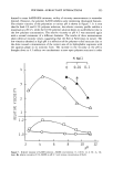
254 JOURNAL OF THE SOCIETY OF COSMETIC CHEMISTS needed for the binding of a reactive hapten to appropriate cells and proteins within skin, and the recruitment and communication with responding T-lymphocytes and other cells. Under physiological conditions, LC continuously present skin-contacting antigens to the T-cells. In this way the skin immune system is continuously made aware of its antigenic microenvironment. Small abrasions and erosions of the skin barrier that nor- mally occur at the microscopic level result in intraepidermal injections of antigenic materials. Other water-soluble compounds will penetrate into the spinal layer of the epidermis and be taken up by LC. When other macrophages fail to clear these antigens, they are processed by the continuously boosted T-cells without any clinical symptoms or signs (2). A large amount of evidence supports the hypothesis that LC are responsible for the critical step of antigen presentation during induction of CH in the skin (13). This evidence includes both circumstantial observations made in vivo and direct observations in experimental animals (3). First, LC are located in relatively large numbers at the cutaneous environmental interface where by implication they should be able to act as APC. Second, experimental work with hapten-derivatized skin grafts has shown that the immunological events that are associated with antigen presentation are accom- plished within the skin graft itself rather than by APC provided by recipients (6). The crucial role of LC in this process is implied by the observation that the major source of IgA immunogenicity in the skin resides with IgA-positive LC (8). It is thus most probably the LC-hapten complex that is involved in presentation of hapten to paracor- tical T-cells. These become activated and are induced to proliferate and differentiate. Thy-1 + DENDRITIC EPIDERMAL CELLS Relatively recently, concepts of cutaneous immunology have been enlarged with the identification in mouse epidermis of a second dendritic cell population, one that ex- presses larger amounts of the cell surface Thy-l-glycoprotein. This cell, termed the Thy-1 + dendritic epidermal cell (Thy-1 + DEC) resembles LC by its marrow derivation (3,9-11), but its expression of Thy-1 antigen suggests an association with T-lympho- cytes or natural killer cells. They resemble primitive T-lymphocytes and may function as epidermal suppressor cells. The functional role of Thy-1 + DEC in humans is un- known, but because it is presumed to be of T-lymphocyte lineage, it is believed to play some role in the cutaneous immune response. KERATINOCYTES, INTERLEUKINS, ETAF Recent work has also demonstrated that the initiation of cellular proliferative responses requires not only physical contact between an antigen-presenting cell (APC) and re- sponding lymphocytes but also the elaboration of a soluble protein, or a "second signal." In the case of T-lymphocyte activation, this second signal is provided by the cytokine interleukin-1 (IL-1), a protein that normally produces the same APC (macro- phage) that is responsible for antigen presentation (12). There is evidence to suggest that epidermal LC produce the monokine IL-1 and present antigens to T-cells. These
Purchased for the exclusive use of nofirst nolast (unknown)
From: SCC Media Library & Resource Center (library.scconline.org)