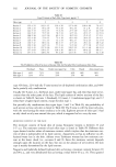
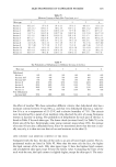
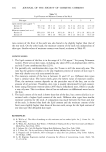
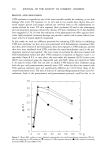
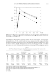
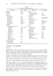
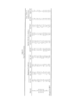
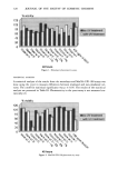
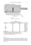
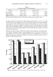
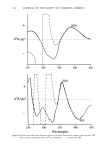
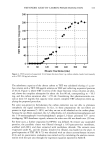
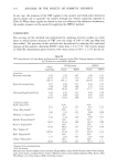
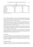
322 JOURNAL OF THE SOCIETY OF COSMETIC CHEMISTS Although not statistically significant, directional trends suggest Material C and Mater- ial E at 0.01% and 0.03% were phototoxic. At 0.05%, however, Material E was cyto- toxic, and reliable phototoxicity measurements were not possible. These findings are consistent with trends seen in the MatTek EPI-100 system. Materials C and E (at 3%) were also phototoxic in the yeast assay. Since no stratum corneum is present to restrict test material contact with cells, the monolayer method is a sensitive means of assessing phototoxicity. In addition, since mammalian cells are used, phototoxicity of the test compound and potential metabo- lites can be assessed. Due to the nature of the test system, however, materials cannot easily be assayed neat. In this test, cytotoxicity was more of an issue than with the Mat- Tek EPI-100 and yeast methods. Also, the absence of a stratum corneum and barrier function does not allow for a realistic evaluation of phototoxicity on intact skin. While no statistical differences were noted with the materials, directional data correlated with the results in the yeast assay. MatTek EPI- 100 A significant (p • 0.05) increase in toxicity as a result of the 8-MOP positive control was noted in tissue irradiated with UVA, indicating phototoxicity. No significant pho- totoxicity (p • 0.05) was seen in this assay with any test material. Although not signif- icant, directional trends indicate Material C was phototoxic. This directional trend was also noted in the monolayer and yeast assays. YEAST This assay was the benchmark used to evaluate results obtained in the monolayer and MatTek EPI-100 assays. Materials that caused a zone of inhibition of greater than 2 mm were defined as phototoxic (2-4). Phototoxicity was noted with the 8-MOP positive control and Materials C and E (3%) in this assay. The phototoxicity associated with Material C in this assay is consistent with the trends noted in both the MatTek EPI-100 and monolayer systems. Although Material E at 3% showed phototoxicity, the same material was not phototoxic at 1%. The difference between 1% and 3% may be a simple concentration effect. At 5 %, Mate- rial E was cytotoxic, as indicated by the large zone of inhibition surrounding the non- irradiated filter disk, and reliable phototoxicity assessments were not possible. DISCUSSION The results seen across the three test systems were similar. Directional trends indicate that Material C was phototoxic in both the MatTek and monolayer systems. Results with the yeast model confirmed this phototoxicity. The two lowest concentrations of Material E were phototoxic in both the yeast and monolayer assays, but, because of cyto- toxicity, phototoxicity was not noted at the highest concentration. The lack of response with Material E in the MatTek EPI-100 system may be due to the presence of the stra- tum corneum, which serves as a protective barrier and limits material penetration to the underlying epidermis.

NEW PHOTOTOXICITY METHODS 323 Each of these systems is useful, depending on the type of application and specific needs. The MatTek EPI-100 system is the most sophisticated system tested, as it is con- structed to be similar to human skin, including a well-defined stratum corneum and barrier function. Because the MatTek system is constructed to be similar to human skin, it is considered to most closely mimic in viv0 test conditions. As the monolayer system is limited by the fact that (a) materials cannot easily be tested neat, (b) there is no pro- tective stratum corneum, and (c) the variability of monolayer systems is inherently high, it may be most useful as a screening tool for phototoxic effects. Finally, the yeast method is a useful and inexpensive means to assess phototoxicity on a large number of materials prior to more expensive testing. The yeast method, however, does not measure the phototoxic potential of test material metabolites in mammalian cells and tests on a substrate dissimilar from the conditions of use. With a good understanding of the limitations and advantages of the different systems, any one of these assays may be used effectively to screen materials for phototoxic effects. Since statistical measurements are more meaningful, the MatTek EPI-100 shows the most promise in this research as a final test for phototoxicity. The data presented here with the monolayer and MatTek EPI-100 systems indicate a good degree of correlation to data obtained using the published yeast assays. Any lack of correlation may be due to differences in the systems as described previously. Finally, it should be noted that the use of the monolayer and MatTek EPI-100 systems for evaluating the phototoxic potential of metabolites from topical preparations is promising. Evaluating metabolites from oral products, however, is more difficult, due to the increased complexity associated with the metabolism of oral products. REFERENCES (1) J. W. Gould, M. S. Mercurio, and C. A. Elmers, Cutaneous photosensitivity diseases induced by ex- ogenous agents,J. Am. Acad. Dermatol., 33(4), 551-573 (1995). (2) E Daniels, A simple microbiological method for demonstrating phototoxic compunts,J. Invest. Der- matol., 44, 259-267 (1965). (3) E. H. Weinbert and S. T. Spring, The evaluation in vitro of fragrance materials for phototoxic activity, J. Soc. Cosmet. Chem., 32, 303-315 (1981). (4) J. D. DiNardo, B. A. Wolf, W. E. Morris, S. Tenenbaum, and R. W. Schnetzinger, A quantitative in vitro assay for the evaluation of phototoxic potential of topically applied materials: Modification and assessment of low-level activity, J. Soc Cosmet. Chem., 36, 423-433 (1985). (5) P. A. Duffy, A. Bennett, M. Roberts, and O. P. Flint, The prediction ofphototoxic potential using hu- man A431 cells and mouse 3T3 cells, In Vitro Toxico/. New Directions, 7, 327-335 (1989).
Purchased for the exclusive use of nofirst nolast (unknown)
From: SCC Media Library & Resource Center (library.scconline.org)