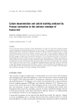
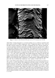
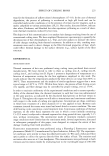
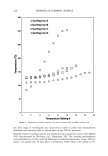
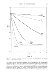
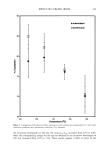
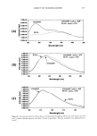
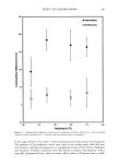
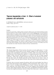
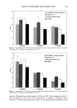
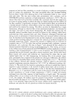
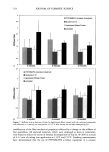
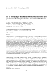
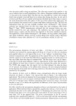
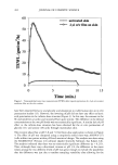
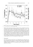
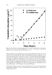
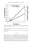
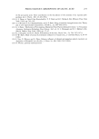
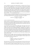
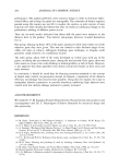
262 JOURNAL OF COSMETIC SCIENCE 6O I untreated skin 2 gl o/w film on skin 5O '•30 • 20 lO 0 5 10 15 Time (min.) Figure 1. Transepidermal water loss measurements (TEWL) after topical application of a 2-pl oil-in-water emulsion film on the skin surface. have been reported for lactic and glycolic acid absorption to viable human skin in i, vitro penetration studies (13). However, the lowering of pH did not have any effect on lactic acid penetration in the infinite-dose situation (Figure 3). In this case, the amount in the SC and epidermis at acidic and neutral pH are quite similar. The difference in the dermal concentrations at the two pH levels was not statistically significant. A similar lack ofpH effect in the infinite-dose situation has been observed for the i, vitro permeation of glycolic (11) and amino (18) acids through mammalian skin. The receptor phase flux at pH 3.8 and 7.0 for finite-dose application is shown in Figure 4. The effect of pH was compared using a completely nested three-way ANOVA (17) (cells within time points within pH level) statistical design. The analysis was done using the SIGMASTAT (Version 2.0) software (Jandel Scientific Software, San Rafael, CA). The analysis indicated that there was no statistically significant difference (p = 0.213). Thus, although there was a directional increase at pH 3.8, the difference in the mean values among the two different levels of pH was great enough to exclude the possibility that the difference was just due to random sampling variability after allowing for the

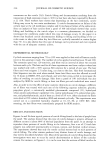
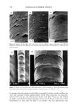
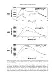
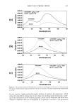
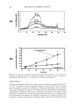
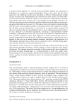
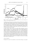
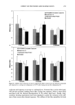
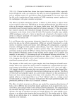
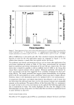
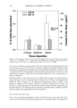
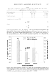
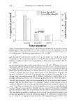
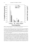
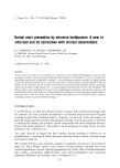
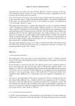
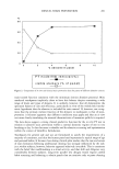
PERCUTANEOUS ABSORPTION OF LACTIC ACID 263 20 * p0.01 pH 3.8 • pH ?.0 T - 40 E 30 ::t. 20 '• 10 =• o o Corneum Epidermis Dermis Tissue deposition Figure 2. Tissue deposition of lactic acid six hours after application of a 2-pl finite-dose o/w emulsion film at pH 3.8 and pH 7.0 (n = 7). Values are mean (n: 7) _+ standard error of mean (SEM). The cumulative receptor penetration at six hours at pH 3.8 is 0.3% -+ 0.1 SEM and that at pH 7.0 is 0.2% _+ 0.0 SEM. effects of differences in time points and cells. The cumulative receptor flux profiles as a function of time for the two application modes are compared in Figure 5. In the infinite-dose situation, a steady state was reached within two hours. The epidermal and dermal concentrations of lactic acid six hours after application of an oil-in-water emulsion are shown in Table II. The tissue concentrations are calculated based on epidermal and dermal thicknesses of 48 and 448 pm, respectively. The data shows that it should be possible to deliver mMolar level of AHA in the living skin tissues from a typical cosmetic rub-on product. Enhanced epidermal cell turnover by lactic and glycolic acid has been suggested (9) as a possible mechanism for their anti- aging efficacy. The results presented here suggest greater bioavailability of ot-hydroxy acids in the SC and epidermis at acidic conditions, which may lead to higher efficacy. However, these in vitro observations need to be validated with in vivo measurements. A comparison of the finite- and infinite-dose delivery data (Figures 2 and 3) shows that the finite-dose film delivered more lactic acid to the stratum corneum and comparable amounts to the epidermis. The greater efficacy of the finite-dose film is a consequence of a rapid increase in the active concentration in the applied film following application due to evaporation of water. The results highlight the fact that the changes in the thermodynamic activity of the active in a finite-dose film can often be a significant factor in active delivery to skin (19). EFFECT OF PROPYLENE GLYCOL The effect of 5% propylene glycol (PG) as a penetration enhancer for lactic acid from
Purchased for the exclusive use of nofirst nolast (unknown)
From: SCC Media Library & Resource Center (library.scconline.org)