j. Cosmet. sci., 52, 155-161 (May/June 2001) Direct evidence for bioconversion of vitamin E acetate into vitamin E: An ex vivo study in viable human skin W. BASCHONG, C. ARTMANN, D. HUEGLIN, and J. ROEDING, M. E. Maeller Institute at the Biozentram, University of Basel, Klingdbergstrasse 5 O- 70, CH-4056 Basel, Switzerland (W. B. ), PhaCos GmbH, Manic& Germany (C.A.), Ciba Specialty Chemicah, P.O. Box 1266, D-79630 Grenzach-Wyhlen, Germany (D.H.), and Mica Prodacts GmbH, Zodlinplatz 4, D-79410 Badenweiler, Germany (J, R. ). Accepted for pablication March 15, 2001. Synopsis For better stability, vitamin E is commonly used as the non-active esterified pro-drug. Such esters are postulated to be hydrolyzed to the free active forrn by skin-related esterases. So far, successful conversion of esterified vitamin E to free vitamin E (tocopherol) has been mainly delineated from observed biological effects. Quantitative evidence in human skin is poor. In vitro and in vivo studies on human and animal skin have proved ambiguous. Formulation-based effects may have added to this controversy. In the present study, comparable amounts of vitamin E acetate (i) in oil (Mygliol-812N), (ii) surfactant- solubilized in water, (iii) encapsulated in liposomes, or (iv) encapsulated in Nanotopes TM were applied to human skin mounted in modified Franz-perfusion chambers that permit emulation of both open or occlusive conditions. The distribution of vitamin E ..... z (vitarnin E acetate +vitarnin E) was assessed on the skin surface, in the horny layers, and in the underlying skin by high-pressure liquid chromatography (HPLC), with a recovery higher than 90%. Vitamin E acetate in Mygliol deposited exclusively on the surface and in the stratum comeurn. In contrast, solubilized or encapsulated vitamin E acetate deposited also in the underlying skin. Nanotopes TM performed best, followed by liposomes and solubilized vitamin E acetate. Non-occlusive application favored deposition in the skin relative to occlusive application. Conversion of vitamin E acetate to vitamin E was not observed on the skin • ,trace or in the horny layers, while in the underlying skin up to 50% of the vitamin E .... • was deacetylated. INTRODUCTION Together with vitamin C, vitamin E is the most common non-enzymatic radical scav- enger involved in protecting living tissue against oxidative stress and radical damage. Incidentally, the human epidermis contains much less vitamin E than other human tissue (1-4). Indeed, topical supplementation of the skin with vitamin E has led to a Address correspondence to W. Baschong. 155

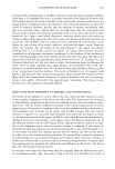
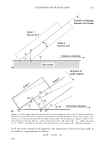
156 JOURNAL OF COSMETIC SCIENCE range of cosmetic effects, such as skin smoothening, moisturizing, prevention of pre- mature skin-aging, suppression of UV-induced erythema, or improved wound healing (4). Vitamin E is readily oxidized when exposed to atmospheric conditions or light (5). In consequence, free vitamin E is only rarely used in cosmetic formulations. It is replaced by stable esterified pro-drugs, usually vitamin E acetate (6). However, such esters are biologically inactive. If applied topically, vitamin E acetate has been reported to be hydrolyzed in the skin to free vitamin E by esterases (7-9). Yet, despite the many reports documenting their biological activity and implying a conversion of the pro-drug, direct quantitative evidence for a conversion of vitamin E acetate to vitamin E in skin is poor and ambiguous (10,11). Vitamin E acetate and vitamin E are both lipophilic compounds. They dissolve in the oil phase of a formula, from where they are expected to be poorly liberated. To increase bio-availability they are commonly formulated in the water phase of a cosmetic formula by means of solubilizers or by specific carriers (6). In the present study, we compared the penetration behavior of vitamin E acetate under non-occlusive and occlusive conditions. In turn, the pro-drug was either dissolved in oil, solubilized in water, or encapsulated in two different carrier systems, i.e., liposomes or Nanotopes TM, the latter being detergent-resistant unilamellar vesicles with a diameter of 20-40 nm (12). The amount of both vitamin E acetate and vitamin E was quantified in three compartments: at the skin surface, in the horny layers, and within the underlying skin. MATERIALS AND METHODS SKIN BIOPSIES Human skin was obtained after informed consent from otherwise healthy donors having undergone plastic surgery for stomach reduction. Upon excision, the skin flaps (ellip- soid, 5 x 20 cm) were surgically liberated of the adhering subcutaneous fat layer, cut to the size of the penetration chamber, and immediately stored in PBS (phosphate-buffered saline, pH 7.2-7.4) at 36øC. TEST FORMULATIONS Final concentrations of 2% vitamin E acetate were dissolved in Mygliol-812N (caprylic/ capric acid triglyceride Condea Chemie GmbH, [Brunsbiittel, Germany]), solubilized in water by Solubilisant-LRI (PPG-26-buteth-26 and PEG-40 hydrogenated castor oil LCW-Wackherr [Saint Ouen L'Aum6ne, France]), encapsulated in liposomes made from soybean phosphatidyl choline with an average mean diameter of 200 nm (Mica Products GmbH [Badenweiler, Germany]), or encapsulated in Nanotopes TM with a mean diam- eter of about 25 nm (Tinoderm E, Ciba Specialty Chemicals [Basel, Switzerland]). PENETRATION Skin flaps were mounted within 1 h after excision in Franz-like perfusion chambers according to Maibach (13), which had been modified as described (14). The skin sup-
Purchased for the exclusive use of nofirst nolast (unknown)
From: SCC Media Library & Resource Center (library.scconline.org)