PENETRABILITY OF OILS IN HAIR 171 HAIR TREATMENTS The oils were used at a level of 0.2 ml/2.5-3 g tresses. The drops of oil were placed on hair swatches and were spread onto the hair fibers with a fine-tooth comb. The samples were stored overnight, and then the oil remaining on the surface was washed with a 20% solution of sodium laureth sulfate and the swatches were rinsed thoroughly, air-dried, and stored at room temperature. Control samples were treated in a similar way, except for treatment with the oils. ANALYTICAL TECHNIQUE: CONCEPT AND BASIC PRINCIPAL The gallium gun emits a pulsed primary ion beam (accelerating voltage of 25 kV). The primary ions impact/bombard the sample surface and ionize atomic species or small fragments of low- and high-molecular-weight molecules. These ionized species, referred to as secondary ions, are highly mobile and volatile, and become easily extracted by an extraction plate and propelled at high speed into a 2-m-long flight tube. A detector at the end of the flight tube detects and records the secondary ions as they arrive at the end of the tube. The velocity (kinetic energy) of the secondary ions depends on their mass: Ekin. = 1/2 mv 2 The smaller the ionized species, the greater their velocity: Velocity = (length of the tube)/(time-of-fiight in the tube) Typical primary ion doses used in this work were on the order of 10 •2 ions/cm 2 for the analysis. This assures that the data is collected within the static limit, i.e., less than 1% of a monolayer was sputtered. Thus, all molecular fragments are indicative of species existing on the surfaces (along the length and in the cross section of the fiber) under investigation prior to analysis. Under these conditions, the sampling depth of TOF SIMS is only -1 monolayer for molecular fragment ions and 1-3 monolayers for atomic species. Since the sampling depth of TOF SIMS is only approximately one molecular layer, only the low-molecular-weight, highly mobile, surface-active components are detected. The higher-molecular-weight compounds are more difficult, if not impossible, to ionize with the 69Ga+ liquid metal ion gun. Therefore, one has to look at the low-molecular- weight fragments of the high-molecular-weight compounds. Detecting the fragments, in turn, is indicative of the presence of high-molecular-weight compounds. Positive and negative mass spectra are plotted as the number of secondary ions detected (y-axis, counts) versus the mass-to-charge ratio of the ions (x-axis, m/z). Instrumental conditions. The work was done at a local surface analytical laboratory, under contract. The specific analytical conditions and instrumentation used for this work are listed below in detail: Instrumentation Primary ion beam Primary beam voltage Primary ion current (DC) Nominal analysis region Charge neutralization Post acceleration Masses blanked Energy filter/Contrast diaphragm Physical Electronics, PHI TFS-2000 69GA+ liquid metal ion beam 25 kV 600 pA (80 pm) 2 yes 8000 V None no/no

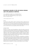
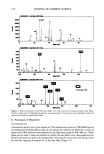
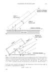
172 JOURNAL OF COSMETIC SCIENCE SAMPLE PREPARATION FOR TOF-SIMS Oils. Small amounts of the pure coconut and mineral oils were deposited on clean silicon wafers at ambient temperature to establish the characteristic positive and negative ions from the mass spectra of the oils for their mapping in the hair cross sections. Untreated and oil-treated hair fibers, The untreated (control) and oil-treated hair fibers were cross-sectioned with a clean stainless steel blade and mounted in small holders with the cross sections facing the spectrometer. OBTAINING ION MASS SPECTRA: ESTABLISHING CHARACTERISTIC POSITIVE/NEGATIVE IONS Positive and negative static TOF SIMS mass spectra were acquired from several locations on each of the oils and from the "interior" cross-sectioned surface of the untreated and oil-treated fibers. In this study, the raw spectra from the pure oils and the fiber "interior," that is, from the surface of the cross-sectioned untreated and oil-treated fibers, were collected and compared. This was done to establish the characteristic positive and negative ions of the pure oils, and to "spectrally" establish their absence or presence in the untreated (control) and oil-treated hair fibers. Since the goal of this study was to establish penetration of the oils into the fiber interior, special attention was directed to mapping the presence of these compounds within the fiber cross section. RESULTS AND DISCUSSION I. Penetration of Coconut Oil ION MASS SPECTRA Coconut oil on a silicon wafir. As the first step, the positive and negative characteristic ions of pure coconut oil, resulting from ion bombardment, have to be established. It is shown that the positive and negative TOF-SIMS spectra of coconut oil contain several charac- teristic ions that can serve as markers for the coconut oil within the hair fibers. Characteristic positive ions of pure coconut oil. Characteristic positive ions are at 127, 155, 171,183,211,257,411,439, and 467 m/z. The positive ion at 127 (126.67) m/z may be the best ion for the imaging of coconut oil, since it is intense and is likely to be free of mass interference. Figure 2 shows the mass spectra of the characteristic positive ions for coconut oil. Characteristic negative ions of pure coconut oil. Characteristic negative ions of pure coconut oil are at 41, 58, 71, 143, 171, 199, and 227 m/z. The individual peaks have not been identified, but many are due to fatty acids such as lauric and oleic acids. Obviously, some of these may be resulting from mixed triglycerides present in minor quantities in coconut oil. Because the coconut oil forms strong positive ions, it tends to form weak negative ions. Therefore, the positive ion at 127 (126.67) m/z will be used for imaging coconut oil in the cross section of the hair fiber (see Figure 3 for the mass spectra of the negative ions for coconut oil).
Purchased for the exclusive use of nofirst nolast (unknown)
From: SCC Media Library & Resource Center (library.scconline.org)