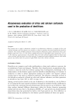
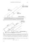
EXTENSION OF HUMAN HAIR 187 by extension of the main shaft. The points on Figure 1 of the measurements of scale angle versus strain obtained by Guiolet eta/. (2) suggest that up to a strain of about 8%, with the fibers at an environmental relative humidity of 35%, there is no slippage between scales, and that above 10% slippage plus some detachment has occurred. This also concurs with the observation in Table 2 of Guiolet's paper (2) that the transparency of the hair is constant up to 7.5% strain and then drops rapidly with further extension. This change would be brought about by an air film forming between overlapping scales along the surface of detachment. These suggestions generally concur with the observations of Gamez-Garcia (3) that the lower the moisture content of the hair fiber under test the lower the strain level at which detachment of the scale structure is initiated. The latter also noted that the lifting of the scales showed no presence of endocuticular debris at their internal surfaces. As suggested elsewhere (4), although the endocuticle has the least concentration of cystine cross-links of the layers in the cuticle cell and the highest swelling in water, it is the upper-p-layer in the cell membrane complex (CMC) that provides the possibility of specific mechanical weakness (see Figure 2). Further discussion on the nature of this p-layer will follow. Ruetsch and Weigmann (5) also have examined the damage to both human hair and wool cuticles by fiber extension to 30-35% strain. In the case of wool fibers, although Hair surface Upper 13-1ayer• i i i i I I I I !1•1 II Exocuticle '''''' (high S) J• Endocuticle (low S) Inner layer 8 layer ••w^•• Lower I• layer "*'--- Upper I•-Iayer i i ill i i i i i i i i i i i i i i ,, ,,,,,, , .............. ,,,,',,,• Exocuticle Iiiii I Iii I I I I I I I Iii I I I I I (a) ' ' "'' ' ' ' ' "'' '"" '''• ' ' ' '"i"'"'"'"' ' ' (highS) Hair surface -. a Upper I• layer "A" I •Ya•re• (nhl gehX•s• uticle- •. / Distortion of endocuticle Endocuticle •/ '••' X upper I• layer "A" layer anhd exocuticle layer ( igh S) Endocuticle • //J Figure 2. A sectional diagram of two overlapping scales showing in 2a details (see text) of the different layers of the scale structure. In 2b are shown the proposed distortion of the low cross-linked endocuticle when the hair fiber is extended, resulting in opposing relative stresses (a and b) in the two scales. At the endocuticle edge (X) these stresses result in shear forces, which may lead to scale lifting.

188 JOURNAL OF COSMETIC SCIENCE no scale lifting was observed, they still claim that failure in the intercellular cement between cuticle cells occurs, with the relative sliding of the scales, in response to the extension of the fibers. THE CUTICLE CELL The cuticle cell surface (4,6), as indicated in the sectional diagram (Figure 2), consists of a thin hydrophobic layer (upper-[B-layer) beneath which are the two layers "A" and exocuticle. Both these latter layers contain a high degree of disulfide bonding, resulting in mechanically inextensible structures low in swelling when placed in a wet environ- ment. The endocuticle layer beneath these rigid structures is low in disulfide cross- linking, resulting in a mechanically more extensible structure capable of a high degree of water uptake. This great difference in water uptake by the "A" and the exocuticle layers as against the endocuticle has been shown by Robinson (7) to produce the pronounced projection of scales in unextended wet wool fibers. This scale projection in the wet wool fibers is the prime cause of "felting" entanglement in wool fiber masses during washing in water. For extension of hair fibers under ambient conditions to 30-35% strain, Ruetsch and Weigmann (5) observed from autofluorescence and scanning electron microscopic stud- ies the cuticular damage and failure at the scale edges, which result in scale lifting. The difference in the stiffness of the "A" and exocuticle layers as against the considerably less stiff endocuticle layer results, during the longitudinal extension of a hair fiber, in a major component of the distortion occurring in the endocuticle (see Figure 2b). On the basis of this result and the observation by Swift (8) that material observed with scale lifting is associated with endocuticular debris, Ruetsch and Weigmann (5) concluded that endocuticular failure precedes scale edge lifting. This conclusion from later observation by Swift (4) and others (3) must be re-examined. Swift (4) noted the failure mechanically of the cell membrane complex (CMC) along the upper-[B-layer. As mentioned earlier, Gamez-Garcia (3) observed no endocuticular debris at internal surfaces of detached scale structure. The possible involvement of the hydrophobic upper- [B-layer, together with its surface of 18-methyleicosanoic acid (18-MEA) in the failure of the cementing of overlapping scale structures and its involvement in standard permanent setting procedures of human hair in salons, is next discussed. THE UPPER-[•-LAYER OF THE CUTICULAR CMC Jones and Rivett (6) have proposed a model for the upper-[B-layer of cuticular CMC consisting of a fatty acid monolayer, approximately 3-nm thick, attached by thio-ester linkages to a proteolipid membrane of approximately the same thickness (Figure 2a). The proteolipid layer in turn is attached to the "A" layer of the exocuticle of the cuticle cell. The fatty acid is the 18-MEA, with the straight chains forming a parallel array of ordered structures terminating in a short branched chain. This parallel chain array is at right angles to the proteolipid membrane, with the branched terminating chain on the surface providing mobility to the fatty acid monolayer. The evidence (9) provided for this increased mobility is the demonstrated lowering of the melting point from 77øC for eicosanoic acid to 56øC for 18-MEA by the introduction of the methyl group. The
Purchased for the exclusive use of nofirst nolast (unknown)
From: SCC Media Library & Resource Center (library.scconline.org)