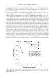
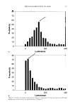
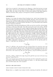
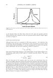
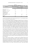
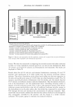
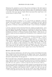
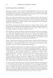
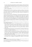
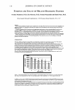
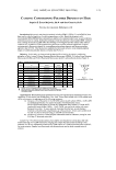
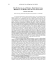
2003 ANNUAL SCIENTIFIC MEETING 120 100 .C' · 80 ·.::, ;;l � 60 :1 40 � 20 0 0J/cm2 2J/cm2 6J/cm2 12J/cm2 UVA dose Fig. I: Effect of UV A exposure on the in vitro catalase activity of EUK-134 (Eukarion, Inc) and catalase from bovine liver (Sigma) Another interesting avenue for the enhancement of the cellular protection mechanisms is to induce the expression of various Heat Shock Proteins which have the properties not only to protect specific UV sensitive enzymes such as catalase, but have been shown to refold the proper terti ary structure of damaged proteins, resulting in a restoration of the activity. Finally, increasing the cell's own energy reserve via topical application of Creatine has been shown to enhance cellular protection mechanisms in UV exposed skin cells, resulting in a significant reduction of both mitochondrial and nuclear DNA damage as well as a reduction of sunburn cell formation. These results clearly demonstrate the relevance of increasing the overall cellular metabolism to improve the overall defense capacity against the oxidative stress generated by exposure to the environment Conclusion These observations strongly support the need for development of a multi-branched technology to provide the necessary protection to cells exposed to the oxidative damages generated by an environment which has evolved dramatically over the last 100 years and which is creating a significant burden to mammalian cells. The development of an optimal protection technology cannot be based exclusively on a simple combination of efficacious sunscreens alone, but should provide a well balanced combination of sunscreens, antioxidants, anti-inflammatory agents, together with ingredients that will enhance the cells' own ability to protect themselves from various environmental insults. We do believe that the cosmetic industry is playing an important role in providing consumers with the best protection available today, as clearly mammaJian cells have not been able to adapt their protective mechanisms to the additional oxidative stress which results from the increase in environmental pollution all over the world. 135

136 JOURNAL OF COSMETIC SCIENCE THE EXTRACELLULAR MATRIX- FROM STRUCTURAL RESILIENCE TO MODULATION OF CELL FUNCTIONS Sanford R. Simon, Ph.D. State University of New York at Stony Brook, Stony Brook, NY 11794-8691 The extracellular matrix (ECM) is classically regarded as having a role in maintaining structural integrity of connective tissue. Two components of the interstitial ECM, collagen and elastin, play especially prominent roles in establishing the balance between rigidity and elasticity which characterizes normal connective tissues. When these proteins are serving as structural elements in the interstitium, they exist predominantly as fibers and are characterized by insolubility and resistance to many proteolytic enzymes. Infiltration of the interstitium by inflammatory cells, especially neutrophils and mononuclear phagocytes (monocytes and macrophages) is traditionally implicated as a critical step in establishing a milieu in which specialized proteinases are released, leading to degradation of collagen and elastin fibers. A net loss of elastic fibers due to chronic degradation is considered to be characteristic of normal ageing of skin, while UV damage to the skin is associated with an accumulation of elastin-like material which has been postulated to result from exaggerated biosynthetic activity triggered by peptides released from inflammatory destruction of normal collagen and elastin fibers. Thus, proteolytic degradation of collagen and elastin has been considered to lead to undesirable changes in the characteristics of skin, resulting either in abnormal thinning or thickening. The proteinases implicated in these changes have been purified and extensively studied as soluble enzymes, and considerable efforts have been directed towards characterizing their endogenous inhibitors as well as developing synthetic inhibitors to supplement the endogenous antiproteinases. This model of degradation of purely structural proteins by soluble enzymes released by inflammatory cells has proved to be oversimplified. In this presentation, we consider some of the results which have emerged from many laboratories indicating that the functional roles of compoµents of the ECM go beyond maintenance of rigidity and elasticity. Moreover, inflammatory proteinases may function in highly specialized microenvironments, such as the inflammatory cell surface, and may have physiologically relevant targets beyond the principal structural elements. Thus, attempts to modulate the activity of these proteinases may have consequences beyond changes in collagen and elastin fiber structure. The Roles of ECM-Associated Matricellular Proteins The ECM which separates epithelial and stromal components, as seen at the dermal-epidermal junction, is referred to as basement membrane (BM), and has unique components, including nonfibrillar collagens (types IV, XV, and XVIII) as well as a variety of bridging elements such as laminin, ent':lctin (nidogen), and heparan sulfate proteoglycans. These components interact not only with each other but also with epithelial and stromal cells. More recently, a number of additional proteins have been characterized which appear to function primarily as cell regulatory molecules rather than structural components. These molecules include thrombospondins, SPARC or osteonectin, osteopontin, and tenascins. With the notable exception of osteopontin, which enhances adhesion of cells to BM through integrins, these so-called matricellular proteins typically reduce the adhesion of cells to BM and may facilitate their migration. Moreover, thrombospodin-2 appears to play an important role in clearing the BM of one of the important matrix-degrading proteinases, MMP-2 or gelatinase A, by binding to the enzyme and facilitating its internalization. Several matricellular proteins regulate such cellular events as proliferation and responsiveness to growth factors, apoptosis, and angiogenesis. Virtually all the matricellular proteins are targets of leukocyte elastase, the serine proteinase released from the azurophil granules of neutrophils. In addition, the combined activities of elastase and the matrix metalloproteinases (MMPs) released by neutrophils and macrophages can degrade types IV and XVIII collagen to liberate proteolytic fragments such as endostatin and tumistatin, which bind to integrins on cell surfaces, resulting in significant anti angiogenic activities. The consequences of pathologically elevated levels of the inflammatory cell-derived proteinases or of abrogation of proteolytic activity by injudicious use of exogenous antiproteinases on the multiple functional roles of the matricellular proteins have not yet been fully explored. Protei11ase-A11tiprotei11ase Imbalance in ECM Damage One of the more challenging aspects of attempting to intervene in preventing excessive damage to ECM through the use of proteinase inhibitors relates to the interactions between the most common proteinases and their endogenous inhibitors. The serine proteinase, leukocyte elastase, is targeted by the endogenous antiproteinases, alpha-1-Proteinase Inhibitor (alpha-I-PI), secretory leukoprotease inhibitor
Purchased for the exclusive use of nofirst nolast (unknown)
From: SCC Media Library & Resource Center (library.scconline.org)