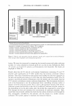
2003 ANNUAL SCIENTIFIC MEETING The Bricks and Mortar Model The permeability barrier of the SC is sometimes modeled as a brick wall with the bricks being the corneocytes with the resistant cell envelopes and keratin microfibrils and the mortar being the intercellular lipids. The mortar is the main barrier to water passing through the SC and lipid soluble molecules are modeled as winding their way through the mortar. The bricks (corneocytes) are 70-80% keratin by dry weight. The keratin is condensed in the form of microfibrils. The keratinocyte cell membrane is replaced by a tough new structure of cross linked protein called the cell envelope. There are lipids covalently attached to the cell envelope on the outer surface and the corneocytes are joined by desmosomes. The keratins in keratinocytes below the SG are in the form of coiled-coiled coils of ex-helix. As the SG cells are transformed into squames the coiled coils aggregate to form 32 chain structures called microfibrils which lay parallel to the surface of the skin and restrict in plane swelling of the squames. Two proteins from the keratohyalin granules, filaggrin and loricrin play key roles in the fonnation of the "bricks". Filaggrin is an acronym for filament aggregating protein. Filaggrin contain a high level of positively charge amino acids and participates in the aggregation of the negatively charged keratin coiled coils. Loricrin is a globular protein that is rich in hydrophobic amino acids and cysteine. It is released from the granules and is crossed-linked to the protein involucrin which is already in the cytoplasm of the cell. The two proteins are cross-linked by the membrane bound enzyme transglutaminase to begin forming the cell envelope. The crosslink is formed between lysine and glutamic acid side chains to form what is known as the isopeptide bond. Eventually the entire cell membrane is replaced by cross-linked protein and lipids (ceramides) are covalently attached to the outer surface of the this new structure which is known as the resistant cell envelope. Keratin microbrils on the inside are also cross-linked to the envelope. The lamellar bodies that appear at the SG contain lipids which are released into the intercellular space as the SC forms. These lipids are glucosyl ceramides, cholesterol, cholesterol esters and long chain fatty acids. In the intercellular space the glucosyl cermides are converted to ceramides and phosopholipids from the original cell membrane are degraded to fatty acids. The SC contains no phospholipids. The lipids in the intercellular spaces arrange themselves into multiple layers. These multilamellar lipids are the mortar of the bricks and mortar model of SC barrier function. References: Marekov and Steinert, Ceramides are bound to structural proteins of the human foreskin epidermal cornified cell envelope, J. Biol Chem. 273, 17763-17770 (1998) Steinert, P.M., Marekov, L.N., Fraser, R.D.B. and Parry, D.A.D. Keratin intermediate filament structure: Crosslinking studies yield quantitative information on molecular dimensions and mechanics of assembly. J Mo/ Bio 230, 436-452. (1993) Steinert and Marekov, Direct evidence that involucrin is a major early isopeptide cross-linked component of the keratinocyte cornified cell envelope. J. Biol Chem. 272:2021-2030(1997) Swartzendruber, D. C., Wertz, P. W., Kitko, D. J., Madison, K. C., & Downing, D. T Molecular models of the intercellular lipid lamellae in mammalian stratum corneum. J Invest Dermatol, 92(2): 251-257. (1989.). Wertz, P. W. & Downing, D. T. Covalently bound omega-hydroxyacylsphingosine in the stratum corneum. Biochim Biophys Acta, 917(1): 108-111. (1987). 133

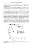
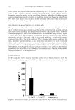
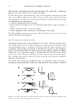
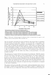
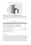
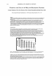
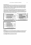
134 JOURNAL OF COSMETIC SCIENCE THE INS AND OUTS OF SKIN PROTECTION TECHNOLOGIES Daniel Maes, Ph.D., D. Collins, H. Corstjens, Ph.D., L. Declercq, Ph.D., R. Foyouzi-Youssefi, Ph.D., D. Gan, L. Hellemans, Ph.D., M. Ingrassia, T. Mammone, Ph.D., M. Matsui, Ph.D., E. Pelle, Ph.D. and I. Sente Estee Lauder Companies, Inc., 12 5 Pine/awn Road, Melville, NY 117 4 7 It is now well accepted that most of the visible signs of aging which appear on the face, are caused by the various factors we are exposed to on a continual basis. As Cosmetic Scientists, we used to believe that most if not all of this damage, visible in the form of lines and wrinkles, abnonnal skin pigmentation, and skin sagginess, were the clinical manifestations of damage resulting from an excessive exposure to the sun. However, it has now become obvious that exposure to solar ultraviolet (UV) and infrared (IR) rays is only partly responsible for the progression of cutaneous damage. Recent evidence suggests that other factors such as smoke from cigarettes, industrial pollution, Ozone which accumulates during the day at ground level, irritants and sensitizers which we get in contact with, and even the psychological stress which we endure in our daily lives play an important role in promoting additional damages to the skin. Clearly, from such an exhaustive list of challenges, it seems likely that the more classical way of protecting the skin with chemical sunscreens will not suffice. Indeed they have the great merit to reduce significantly for some time the damages caused by UVB and UV A to essential bio-molecules, such as DNA lesions as well as oxidized proteins and lipids. But oYer the past 15 years substantial progress has been made by marrying the effect of sunscreens with the activity of topical antioxidants, in order to provide broader and longer lasting protection benefits. During that time period, a few long-term clinical studies have demonstrated tl1e benefits one can expect from the regular treatment of the skin with such molecules. They do however present some challenges to the formulator, as their lack of stability in an emulsion, obviously due to their antioxidant activity, limits sometimes tl1eir usage to low concentrations that do not allow for the complete control of the oxidative damage taking place in the skin. As a consequence, a fairly complex and devastating cascade of events is taking place: First, tl1e oxidafo·e damage to the cell membrane will induce the release of proinflammatory mediators that will ultimately lead to the activation of metalloproteases, capable of gradually degrading the extracellular matrix. The end result will be a loss of integrity, firmness and elasticity of the skin. A second, even more dreadful impact of the oxidative damage to our cells is certainly linked to protein oxidation. Indeed, this process will not only affect the function of essential structural proteins such as collagen, elastin and keratins, but in addition it can lead to a loss of activity for key enzymes in our skin. Such deactivation can have an impact at various levels. It can affect barrier integrity by perturbing the cellular desquamation process, since the stratum comeum chymotriptic enzyme (SCCE) is susceptible to UV-induced oxidative deactivation. It can also compromise the internal antioxidant defense mechanism since catalase, responsible for the reduction of hydrogen peroxide into water, is known to be deactivated by UVA exposure. More recent developments in technology allow us to compensate for such deactivation of the cells' own ability to protect tl1emselves from these oxidative damages. Specific molecules have been synthesized or extracted from plants to reactivate the deficient antioxidant mechanism such as N-Acetyl-Cysteine to replenish the pool of intra-cellular glutathione, or the UVA stable catalase mimetic EUK-134 (Fig. 1), to compensate for the UVA-induced loss of catalase activity.
Purchased for the exclusive use of nofirst nolast (unknown)
From: SCC Media Library & Resource Center (library.scconline.org)