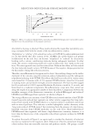
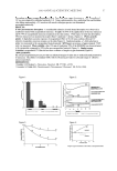
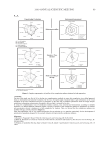
JOURNAL OF COSMETIC SCIENCE 2 (delipidation), as well as formation of acid groups (acidifi cation) of the exposed scale faces, changes the originally hydrophobic nature of the scale faces and gives them hydrophilic properties. Most of the information we have on the nature of lipids on the surface of hair origi- nated from studies on wool. On the basis of persistence of hydrophobicity in wool after solvent extraction and mild scouring, Leeder and Rippon (3) concluded that the lipids left on the surface were covalently bound to the epicuticle. Evans et al. (4) and Kalkbrenner et al. (5) found that the covalently bound lipids can be released from the surface by treatment with potassium tert-Butoxide in tert-butanol. Negri et al. (6) found that chlorination released over 50% of the bound fatty acids, and these were mainly bound by thioester linkages. However, fatty acids bound by ester or amide linkages were cleaved only by hot aqueous treatments. The observation that acidic chlorine water is capable of releasing covalently bound fatty acids from the surface of hair is relevant to human hair, since the hair often encounters chlorine in the water of swimming pools. Wertz and Downing (7) approached the problem of lipids on the surface of human hair from the studies of lipids of mammalian epidermis. Their detailed analytical study of human hair showed that 18 MEA (C-21 ante-iso) forms only 40% of the covalently bound lipids. In the current study we attempt to characterize and quantify changes in the surface chemistry of hair as a function of progressive reduction. Since surface chemistry is important from the point of view of the spreading of hair care products and the friction that affects the feel of hair, it would be important to characterize the surface of such chemically treated hair. As in earlier work (1,2), we again use the same microfl uoromet- ric technique with the help of the cationic fl uorochrome Rhodamine B to detect the change in the surface chemistry of reduced hair. More specifi cally, we attempt to quan- tify and compare the level of photochemically and cosmetic-chemically (oxidation and reduction) induced breakdown of the thioester linkages, removal of the surface lipids (irrespective of their chemical composition), and formation of acid functionalities on the scale faces. In addition to microfl uorometry, we attempt to measure this change in sur- face chemistry by XPS analysis and wettability scanning (8). The effect of delipidation of the hair surface on friction and the positive effect of depositing conditioners on such damaged hair have been presented in an earlier publication (9) and will not be pursued in this communication. The concept of the microfl uorometric approach used in this research is based on the fact that the intact lipid layer resists rapid adsorption of the cationic fl uorochrome and is indicated by the low fl uorescence intensity of the scale faces. On the other hand, removal of the lipid layer, and formation of a large number of acid groups on the exposed scale faces, enhances rapid adsorption of large amounts of cationic fl uorochrome, resulting in the high fl uorescence intensity of the scale faces. Alternate methods, which are highly sensitive to changes in the surface chemistry of the scale faces, are single-fi ber wettability scanning and XPS analysis. Wettability scanning, which is a measure of surface wettability, detects and measures changes in surface chem- istry from hydrophobic to hydrophilic as a result of oxidation or reduction processes. XPS can establish treatment-induced changes in the concentration and chemical state of all detectable elements at a surface depth as shallow as 25Å. We use these techniques to confi rm and support the results obtained by the microfl uorometric approach.

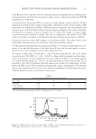
REDUCTION-INDUCED HAIR SURFACE MODIFICATION 3 EXPERIMENTAL MATERIALS AND TECHNIQUES (a) Unaltered hair samples. A tress of 14˝-long, unaltered, European dark brown hair from DeMeo Bros., New York, was used for this study. (b) Hair sample preparation. Eight inches of the root portions of individual hair fi bers were taken and numbered 1 to 30. These root portions were then cut into six equal segments. Starting with the top section of each hair fi ber, the segments were numbered 30, 15, 10, 5, 2, and 0 minutes. (c) Reduction treatment. The numbered segments were then subjected to reduction with ~0.5 M ammonium thioglycolate (TGA at pH 9.4 with ammonium hydroxide) for 30, 15, 10, 5, and 2 minutes, while the bottom section (numbered “0”) served as an unaltered, “not reduced” control. The reduced fi ber segments were thoroughly rinsed in lukewarm run- ning tap water for ten minutes and blotted between paper towels. The fi ber segments were air-dried. (d) Fluorochrome. A 0.020% aqueous solution of the cationic Rhodamine B (CI Basic Vio- let 10), (Aldrich Chemical Co., Milwaukee, WI) was used as labeling agent to highlight, characterize, and quantify the oxidative damage infl icted upon the scale surface (1). (e) Tagging of the hair with Rhodamine B. The untreated and reduced hair fi bers were treated for one minute with 0.020% aqueous Rhodamine B solution, actively rinsed for 15 sec- onds in warm running tap water, blotted between paper towels, blow-dried, and stored in the dark at ambient temperature. (f ) Instrumental settings for microfl uorometric scanning. A Leitz MPV 1.1 microspectropho- tometer (Ernst Leitz Wetzlar, GmbH, Wetzlar, Germany) with a Vertical Ploem Illumi- nator, a microfl uorometry unit, was used for this study. Instrumental settings for the spectral and spatial (cross-sectional) microfl uorometric measurements (scans) of the Rho- damine B-labeled unaltered and “reduced” hair fi bers were as follows: • Green excitation beam: 515-560 nm KP = 580 nm LP = 580 nm • λm: 608 nm (fi lter: 37.5 mm) • Objective: 40 X • Accel. voltage: 1.2 kV • Measuring sensor: (5×60) units2 for spectral and distance (spatial) scans • Scanning speed: 72 μm/s for distance scans (high-speed scans) The dried, RB-tagged hair segments were mounted in parallel on microscope slides for spatial scanning. From the fl uorescence emission spectrum of a Rhodamine B-tagged hair fi ber (1), the wavelength of maximum fl uorescence emission had been established at λm ~ 608 nm. All spatial scans were carried out at this wavelength under identical instrumental settings. (g) Wettability scanning. Single-fi ber wettability scanning was carried out using the Wilhelmy technique (8) (using our TRI/scan apparatus). (h) XPS analysis. XPS analysis of the samples was done at an outside analytical facility for an appropriate number of hair fi bers to assure confi dence and reliability in the obtained results.
Purchased for the exclusive use of nofirst nolast (unknown)
From: SCC Media Library & Resource Center (library.scconline.org)