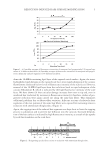
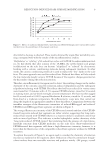
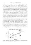
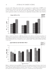
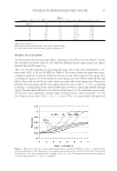
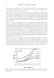
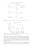
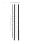
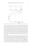
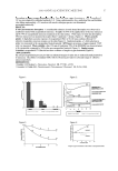
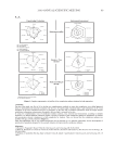
JOURNAL OF COSMETIC SCIENCE 4 RESULTS AND DISCUSSION BACKGROUND In earlier work (9), we characterized and quantifi ed the extent of cuticle cell ablation/ abrasion and complete erosion along the human hair fi ber caused by physical means. We demonstrated, with the help of a fl uorochrome (Rhodamine B in this case), how everyday standard grooming practices severely damage the physical nature of the surface structures (the cuticula) of hair fi bers (9). In other studies (1,2), we attempted to characterize and quantify photochemically and chemically induced oxidative damage to the outer β-layer on the exposed cuticle cell surface. These earlier studies have provided some interesting results, indicating distinctly different phases of hydrolysis-induced 18-MEA scission. The highlights of these studies will be briefl y summarized for the ease of comparing the effects of photochemical and chemical oxidation of earlier studies with the effects of re- duction on the outer β-layer of the exposed scale faces (our current research). (a) Photochemical oxidation. Photochemical oxidation is apparently a “two-phase” process as clearly shown in Figure 1a showing the interfi ber averages (~1200 data points per scan) of progressively UV-exposed segments of 30 different hair fi bers. As can be seen in the plot, there are two distinct phases of photodegradation of the cuticula: (1) short-term light exposure, which is an initiation period of physical changes, especially at the scale edge (as observed in the SEM), preceding lipid removal on the scale faces, and (2) long- term light exposure, during which lipid scission (delipidation) and formation of acid functionalities (sulfonic acid groups) on the scale faces take place. The kinetics of photodegradation, (see Figure 1a), may be explained as follows: The ini- tial and rather constant fl uorescence intensity (FI) of up to 48 hours suggests that this may be an induction period during which photodegradation is suppressed by free-radical generation in the sample. The source of these free radicals could be the ferrous iron in the hair that can generate free radicals by the well known Fenton’s reaction. These free radi- cals (mainly OH and OOH) are very active and mobile and terminate faster than propa- gate free-radical chain reactions. When all the iron is converted to ferric iron, the internal source of free radicals is exhausted. This seems to occur by the end of 48 hours, after which time span the photochemical degradation reaction by direct photolysis of keratin takes over. In addition, free radicals generated by the high-energy photons in combina- tion with water and oxygen may also contribute to overall photolysis. This strongly sug- gests that photochemical oxidation occurs through a free-radical mechanism, leading ultimately to negatively charged cysteic acid groups, which are tagged with RB. (b) Chemical oxidation (bleaching). Bleaching with alkaline hydrogen peroxide, on the other hand, involves thioester hydrolysis at high pH, leading to delipidation, combined with some cystine disulfi de cleavage. Both these reactions lead to formation of cysteic acid at the end, which adsorbs RB. This leads to a monotonic increase in fl uorescence as shown in Figure 1b. CURRENT STUDY: REDUCTION-INDUCED DAMAGE TO THE OUTER β-LAYER OF THE EXPOSED SCALE FACES The present study again uses microfl uorometry to establish how reduction with ammo- nium thioglycolate spontaneously initiates damage to the scale faces by attacking/breaking

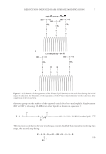
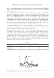
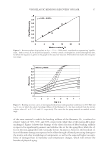
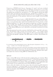
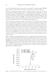
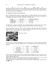
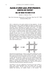
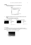
REDUCTION-INDUCED HAIR SURFACE MODIFICATION 5 down the 18-MEA-containing lipid layer of the exposed cuticle surface. Again, the intact hydrophobic lipid domains on the exposed scale faces resist rapid adsorption of the cationic fl uorochrome (indicated by low levels of fl uorescence intensity of the scale faces). However, removal of the 18-MEA lipid layer from the scale faces leads to rapid adsorption of the cationic Rhodamine B, which is indicated by the high fl uorescence intensity of the scale faces. We have shown (9) that cuticular damage increases from root to tip end, even in unaltered hair (indicated by increases in fl uorescence intensity) therefore, studies of pro- gressive reduction were again carried out along the same hair fi bers. This eliminates additional problems due to fi ber-to-fi ber variation. Adjacent sections of relatively short segments of the root portion of the same hair fi bers were exposed for increasing times to reduction with ammonium thioglycolate, (Figure 2). Again, the tagging time of the reduced hair segments was kept short to limit the tagging process to adsorption and to prevent dye diffusion into the cuticula. Increased delipida- tion of the hair surface is indicated by high fl uorescence intensity, as a result of dye uptake by acid functionalities on the scale faces. Figure 1. (a) Interfi ber averages of fl uorescence intensities of unexposed and progressively UV-exposed seg- ments of 30 different hair fi bers. (b) Interfi ber averages of fl uorescence intensities of unexposed and progres- sively chemically oxidized segments of 30 different hair fi bers. Figure 2. Designating specifi c fi ber sections to reduction with ammonium thioglycolate.
Purchased for the exclusive use of nofirst nolast (unknown)
From: SCC Media Library & Resource Center (library.scconline.org)