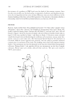
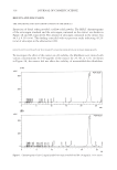
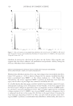
A. INCISUS EXTRACT AND WRINKLE REDUCTION 313 MATERIALS AND METHODS PLANT MATERIAL AND THE EXTRACTION PROCESS The heartwood of A. incisus was collected from Phitsanulok Province, Thailand. The heartwood portion of A. incisus was chipped and dried at 50°C by using a hot-air oven. Then the dried-chipped heartwood was milled into a powder. Five-hundred grams of the A. incisus powder was macerated with 800 ml of diethyl ether (LabScan Asia, Co. Ltd., Bangkok, Thailand) at room temperature for two days, as used previously with slight modifi cation (20). The mixture was fi ltered through a cloth to remove particu- lates, and then the diethyl either was removed by evaporation with a vacuum evapora- tor set at 33°C. The resultant powder was stored in a tight amber glass at −20°C for further studies. QUALITY CONTROL OF THE EXTRACT To control the extract quality of each batch, the content of the artocarpin, a major com- ponent of the A. Incisus heartwood extract, was determined by using isocratic high-per- formance liquid chromatography (HPLC). The artocarpin was provided by Assist Prof. Atawit Somsiri, Faculty of Pharmaceutical Sciences, Naresuan University (25). The HPLC instrument consisted of an SPD-10M10AVP diode array detector and an SCL-10A central unit (Shimadzu Co., Ltd., Kyoto, Japan). An Alltima 250 × 4.60-mm column containing 5 μm of C18 was the stationary phase (Alltech Associates Inc. Corperation, Illinois). The mobile phase was methanol (80 parts) (HPLC grade, LabScan Asia Co. Ltd.) and water (20 parts). The fl ow rate was 1 ml/min and the injection volume was 20 μl. The quantifi - cation of artocarpin was based on peak area at 282 nm. Determinations were performed in triplicate. EFFECTS OF EXTRACT ON THE VIABILITY AND PROLIFERATION OF HUMAN FIBROBLASTS Cells and treatment. Fibroblasts were obtained from a healthy female aged 58 years. Der- mal tissue had been collected aseptically from the nonwrinkled and the wrinkled facial areas situated at the outer corner of the eye. Three-millimeter disks of skin were cut using a biopsy punch. Two to three skin disks were then placed in a 25-cm2 fl ask and subse- quently incubated for 1 h at 37°C with a humid atmosphere containing 5% CO2. After incubation, the tissue disks could well attach on the wall of the culture fl ask. The culture medium consisted of DMEM (PanTM Biotech GmbH, Aidenbach, Germany), 10% FBS (PanTM Biotech GmbH), and 1% of a stock penicillin/streptomycin solution (PanTM Bio- tech GmbH) 3 ml was added to each fl ask. After incubation for three weeks at 37°C with 5% CO2, the fi broblast cells had migrated from the original site. The fi broblast cells were then detached by trypsinization using trypsin-EDTA solution (PanTM Biotech GmbH) and seeded at 1 × 10⁴ cells/cm2 in 75-cm2 fl asks using the same medium. Passage numbers 5 to 7 were used in this study. For the cell treatment procedures, the cell suspension from nonwrinkled or wrinkled skin was transferred from the 75-cm2 fl ask into a 96-well plate at a density of 5×10³ cells/well or a 12-well plate at a density of 4 × 10⁴ cells/well for the cell viability or cell proliferation

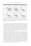
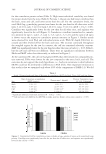
JOURNAL OF COSMETIC SCIENCE 314 studies, respectively. Cells were initially cultured in the culture medium at 37°C for 24 h. Then the medium in each well was replaced with serum-free DMEM containing vari- ous concentrations of the extract (0.5, 1, 2, 5, 10, 20, and 50 μg/ml). Dimethyl sulfoxide (DMSO, 99.5% GC, Sigma-Aldrich, Inc., Missouri) was used to enhance the solubility of the extract in DMEM, and the amount used was not more than 0.1% in the fi nal concen- tration. There were three groups of cultures: (i) serum-free DMEM (untreated group), (ii) serum-free DMEM + DMSO (0.1%, control group), and (iii) serum-free DMEM + extract (treated group). Cell viability and proliferation assay. The activity of mitochondrial dehydrogenases was determined by the 3-(4,5-dimethylthiazol-2-yl)-2,5 diphenyltetrazolium bromide test (MTT, Sigma-Aldrich, Inc.). This test is an alternative method to measurement of the cellular viability as mitochondrial dehydrogenases from living cells are able to convert soluble MTT to an insoluble formazan via a reduction reaction. After incubation under 5% CO2 at 37°C for 24, 48, or 72 h, the cell-free supernatants were removed and replaced with 100 μl of serum-free DMEM. Fifteen microliters of MTT solution (5 mg/ml in PBS) was added to each well. After incubation for 4 h, 100 μl of extraction buffer consisting of 10% w/v sodium dodecyl sulfate (Sigma-Aldrich, Inc.) in 0.5 M N,N-dimethylforma- mide (Sigma-Aldrich, Inc.) was added. The samples were then incubated overnight under 5% CO2 at 37°C. The optical density of the converted dye was measured at 510 nm by using a Labsystems Multiskan RC 96-well microplate reader (Thermo LabSystems, Inc., Massachusetts). Optical density was adjusted to 100% using the untreated cell groups, and the cell viability results were thus expressed as a percentage. The measurements were performed in triplicate. In this study, cell proliferation was determined by counting the number of viable cells in the individual wells by using the trypan blue exclusion test. Cells were treated with the extract or DMSO for 1, 3, 6, or 10 days. The medium was replaced every three days with fresh medium of the appropriate type. The study was performed in triplicate to obtain the average number of viable cells. The appearance of the cells was verifi ed by microscopic examination. Cell cycle analysis. After 72 hr, the proportion of cells in each of the cell cycles, (G1, S, and G2) was determined by staining the cell DNA with propidium iodide (PI). Briefl y, the cells were detached by trypsinization and washed twice with phosphate buffer sa- line (PBS without Ca2+ and Mg2+, PanTM Biotech GmbH) containing 2 mM of diso- dium ethylenadiamine tetracetic acid (Na EDTA). The cells were fi xed overnight with cold absolute ethanol and then stained with a solution containing 5 μl of PI (0.1 mg/ml, Sigma-Aldrich, Inc.), 1 μl of RNAse (1 mg/ml, Sigma-Aldrich, Inc.), and 49 μl of 2 mM Na EDTA in PBS. After 5-min incubation at room temperature in the dark, fl uorescent cells were sorted in a CytomicsTM FC 500 fl ow cytometry system equipped with a 488-nm argon laser (Beckman Coulter, Inc., California). The data were analyzed on the RXP software. EFFECTS OF THE EXTRACT ON PRODUCTION OF TYPE I PROCOLLAGEN AND MATRIX METALLOPROTEINASE-1 (MMP-1) BY HUMAN FIBROBLASTS Cells and treatment. Fibroblasts from cultures of the same explants as above (passage numbers of 5 to 7) were used in these experiments. Before being treated, the cell
Purchased for the exclusive use of nofirst nolast (unknown)
From: SCC Media Library & Resource Center (library.scconline.org)