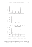
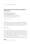
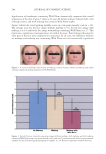
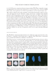
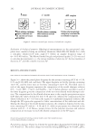
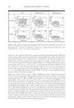
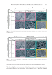
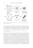
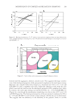
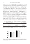
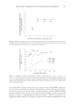
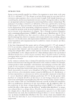
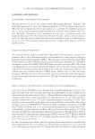
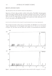
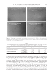
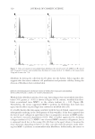
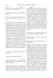
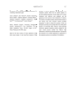
A. INCISUS EXTRACT AND WRINKLE REDUCTION 319 In addition, a cell cycle study of the fi broblasts treated with the extract was conducted by fl ow cytometery. The cell cycle stages in the control (with 0.1% DMSO) and the treated condition (with 50 μg/ml extract) are summarized in Table I, and histograms of the fl ow cytometric data are shown in Figure 5A–D. In comparison to the fi broblasts from wrin- kled skin, the number of cells in the G2 phase of the fi broblasts from nonwrinkled skin was about twofold higher. The extract increased the proportion of wrinkled-skin fi bro- blasts treated to be in the G2 phase by about 2.6-fold. The same trend was found for ex- tract-treated nonwrinkled-skin fi broblasts, but the effect was smaller (1.7-fold). A concomitant increase in cell numbers in the S phase by about 3.6-fold was seen for wrin- kled-skin fi broblasts treated with the extract. This reciprocal relationship between the S and G2 phases indicates that the extract could have increased proliferation of wrinkled-skin Figure 4. Morphology of nonwrinkled-skin fi broblasts (A) treated with 0.1% DMSO or (B) treated with 50 μg/ml of extract and wrinkled-skin fi broblasts (C) treated with 0.1% DMSO or (D) treated with 50 μg/ml of extract for 72 h (at magnifi cation of 10×). Table I Percentage of Fibroblasts from Nonwrinkled and from Wrinkled Skin at Different Stages of the Cell Cycle (G1, S, and G2) after Treatment with 50 μg/ml of A. incisus Extract for Three Days Cell type Treatment G1 (%) S (%) G2 (%) Nonwrinkled-skin fi broblasts 0.1% DMSO 86.59 3.53 9.88 50 μg/ml extract 76.80 5.99 17.21 Wrinkled-skin fi broblasts 0.1% DMSO 89.77 4.66 5.57 50 μg/ml extract 67.94 17.82 14.24

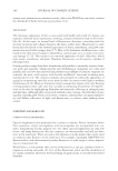
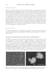
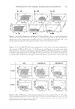
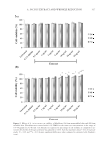
JOURNAL OF COSMETIC SCIENCE 320 fi broblasts by driving the cells from the G1 phase into the S phase. Taken together, this suggests that the extract enhances cell proliferation and promotes viability during the division of fi broblasts from wrinkled skin. EFFECTS OF THE EXTRACT ON PRODUCTION OF TYPE I PROCOLLAGEN AND MATRIX METALLOPROTEINASE-1 (MMP-1) BY HUMAN FIBROBLASTS Wrinkled-skin fi broblasts produced less type I procollagen than nonwrinkled-skin fi bro- blasts (76% greater, p 0.01) as shown in Figure 6A. In contrast, wrinkled-skin fi bro- blasts accumulated more MMP-1 in the culture medium ( p 0.05, Figure 6B). Nevertheless, the extract suppressed MMP-1 synthesis by fi broblasts from both skin types, but the effect on procollagen was confi ned to wrinkled-skin cells. It is widely known that skin aging correlates with the loss of dermal connective tissue, subsequently resulting in the development of skin wrinkles. It has been reported that the loss of type I collagen in aged skin is due to a progressive increase in MMP synthe- sis and hence increased degradation (9,19). This is further aggravated by declining collagen synthesis (14,27). MMP-1 one of several MMP cutaneous collagenases, but it is expressed much less in the young (28–30) than in the aged, where it becomes the more dominant degrading enzyme (31). This coincides with the present study show- ing less MMP-1 synthesis in nonwrinkled-skin fi broblasts. Additionally, the extract at Figure 5. Cell-cycle analysis of nonwrinkled-skin fi broblasts (A) treated with 0.1% DMSO or (B) treated with 50 μg/ml of extract and wrinkled-skin fi broblasts (C) treated with 0.1% DMSO or (D) treated with 50 μg/ml of extract for 72 h.
Purchased for the exclusive use of nofirst nolast (unknown)
From: SCC Media Library & Resource Center (library.scconline.org)