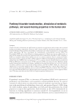
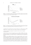
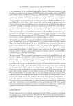
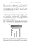
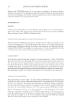
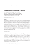
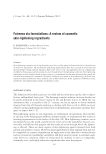
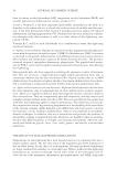
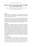
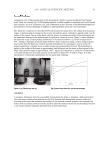
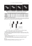
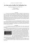
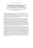
JOURNAL OF COSMETIC SCIENCE 2 topical corticosteroids, has been extensively studied (1,3). Furthermore, its predominant role as a strong anti-irritant and its ability to protect and rebuild the skin barrier through the stimulation of barrier lipid synthesis has been suggested by several studies (4–7). The capacity of PAN to repair the skin barrier has also been postulated as a possible way for PTA to normalize sebum release by strengthening the follicular barrier in persons with oily skin (8). Although several clinical studies on the effi cacy of PAN are available, very little is known on the biochemical mechanism involved. In the context of wound healing, a study on fi broblasts has shown the capacity of PAN to increase protein synthesis and release (9). Interestingly, PAN and PTA have shown the capacity to protect skin from UV-induced erythema (1 G. Dell’Acqua, unpublished data), and data on in vitro and on human skin explants have shown that both PAN and PTA would stimulate the proteins involved into UV-induced oxidation protection and repair (10–12). Taken together, these data suggest that an array of genes could be stimulated by PAN and PTA. Recently, re- search on proliferating fi broblasts in connection with wound healing has shown the acti- vation of genes such as IL-6, IL-8, and HMOX-1 by calcium pantothenate (13), while studies on normal human keratinocytes in connection with an infl ammatory profi le have shown the capacity of PTA in combination with fl avonoids to inhibit a series of pro- infl ammatory genes (7). In order to better understand the underlying molecular mechanism of PTA, we have de- cided to investigate the activation of several biochemical markers in human skin explants when treated with PTA at 2% in an emulsion. We have compared these data with D-panthenol (PAN) in the same condition. We have verifi ed in vivo that PTA penetrates the stratum corneum and that it is de-acetylated in PAN. We have, fi nally, analyzed the wound-healing properties of PTA compared to PAN when applied on the skin by measur- ing the transepidermal water loss (TEWL) of human volunteers who have been subjected to suction blisters. Figure 1. Carbohydrates, fats, and lipid metabolism summary. D-panthenyl triacetate is a derivative of D-panthenol and pantothenic acid, a component of acetyl CoA.

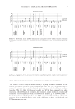
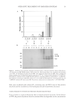
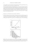
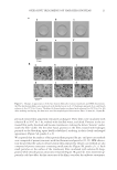
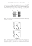
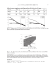
PANTHENYL TRIACETATE TRANSFORMATION 3 MATERIALS AND METHODS SUBSTANCES D-panthenyl triacetate (Induchem AG, Volketswil, Switzerland) and D-panthenol (Dai- ichi Fine Chemical, Takaoka, Japan) were tested diluted in either a water-based gel or in different emulsions. RAMAN SPECTROSCOPY ON THE SKIN OF HUMAN VOLUNTEERS Human volunteers (n = 3) applied three different products on three different areas on the volar side of the forearm: a water-based gel control, a water-based gel containing 3% PTA, and a water-based gel containing 3% PAN. The applied volume of the product was 2 mg/ cm2. The treated areas were measured by confocal Raman microspectroscopy (3510 Skin Composition Analyzer, River Diagnostics BV, Rotterdam, The Netherlands) one hour, fi ve hours, and 24 hours after application. The measurements were taken to a depth of approxi- mately 25 μm in the skin, covering the whole stratum corneum and a small layer (+/- 5 μm) in the viable epidermis. Ten repeated measurements were taken to obtain an average for the heterogeneity of the skin. The results were expressed as milligrams per active/gram of kera- tin versus the skin depth. The results were averaged for all three volunteers. HUMAN SKIN EXPLANTS AND TREATMENT Twelve skin explants were prepared from an abdominal skin biopsy removed in plastic surgery adipose tissue was removed, and the skin was cut off (5 cm2), placed in DMEM medium (10% FCS) into six-well plates, and maintained at 37°C, 5% CO2, humidity controlled. The medium was then replaced by new medium, and different products were topically applied on the epidermis in the plates. The products were a placebo cream, cream containing 2% PAN, and cream containing 2% PTA. The plates were then incu- bated for six or 24 hours. Untreated skin explants were used as controls in parallel. After each incubation period, three punches were performed on each skin explant and washed in phosphate buffer saline (PBS). The epidermal tissue was removed from the punches and immediately frozen at -80°C for further mRNA extraction. QUANTITATIVE RT-PCR ANALYSIS The mRNA of each segment of epidermis was extracted using Tri-Reagent® (Ambion, Austin, TX). The contaminant DNA was removed by treatment with a “DNA-free” sys- tem kit (Ambion), and reverse transcription of mRNA was conducted in the presence of oligo(dT) and Superscript II reverse-transcriptase (Invitrogen, Carlsbad, NM). The mRNA of triplicates was pulled together. Twenty-seven markers of skin metabolism were analyzed (see Table II) by quantitative RT-PCR. A PCR (polymerase chain reaction) was performed in triplicate using the LightCycler® system (Roche Molecular Systems Inc., Pleasanton, USA). WOUND-HEALING DOUBLE-BLIND CLINICAL STUDY The double-blind study was conducted on 40 subjects (female and male) with normal, healthy skin. The 37 subjects who fi nished the study were of ages between 40.3 and 60.8 years
Purchased for the exclusive use of nofirst nolast (unknown)
From: SCC Media Library & Resource Center (library.scconline.org)